Central Areolar Choroidal Dystrophy
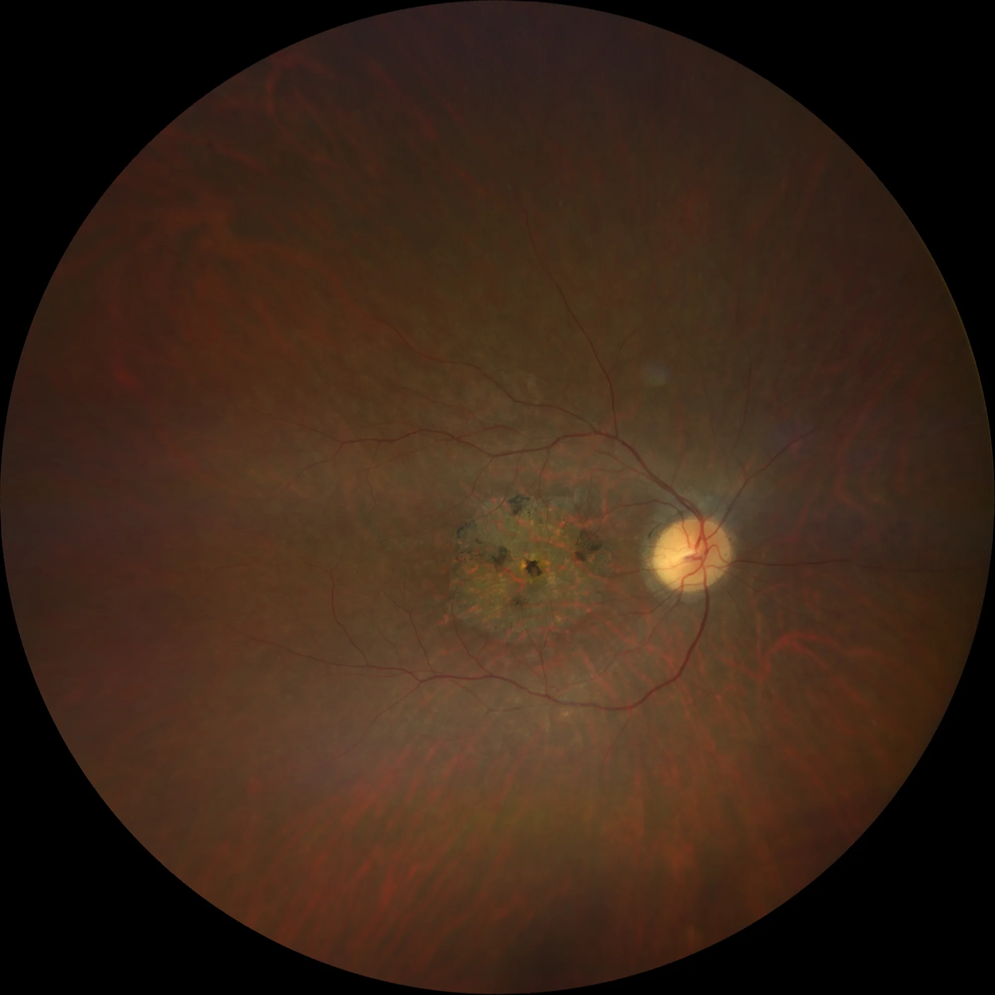
Retinography (Clarus 700, Zeiss): macular atrophy with intralesional and central pigment deposition (A1, A2).
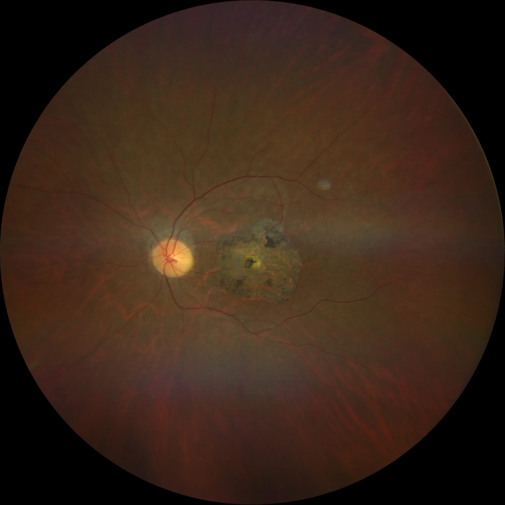
Retinography (Clarus 700, Zeiss): macular atrophy with intralesional and central pigment deposition (A1, A2).
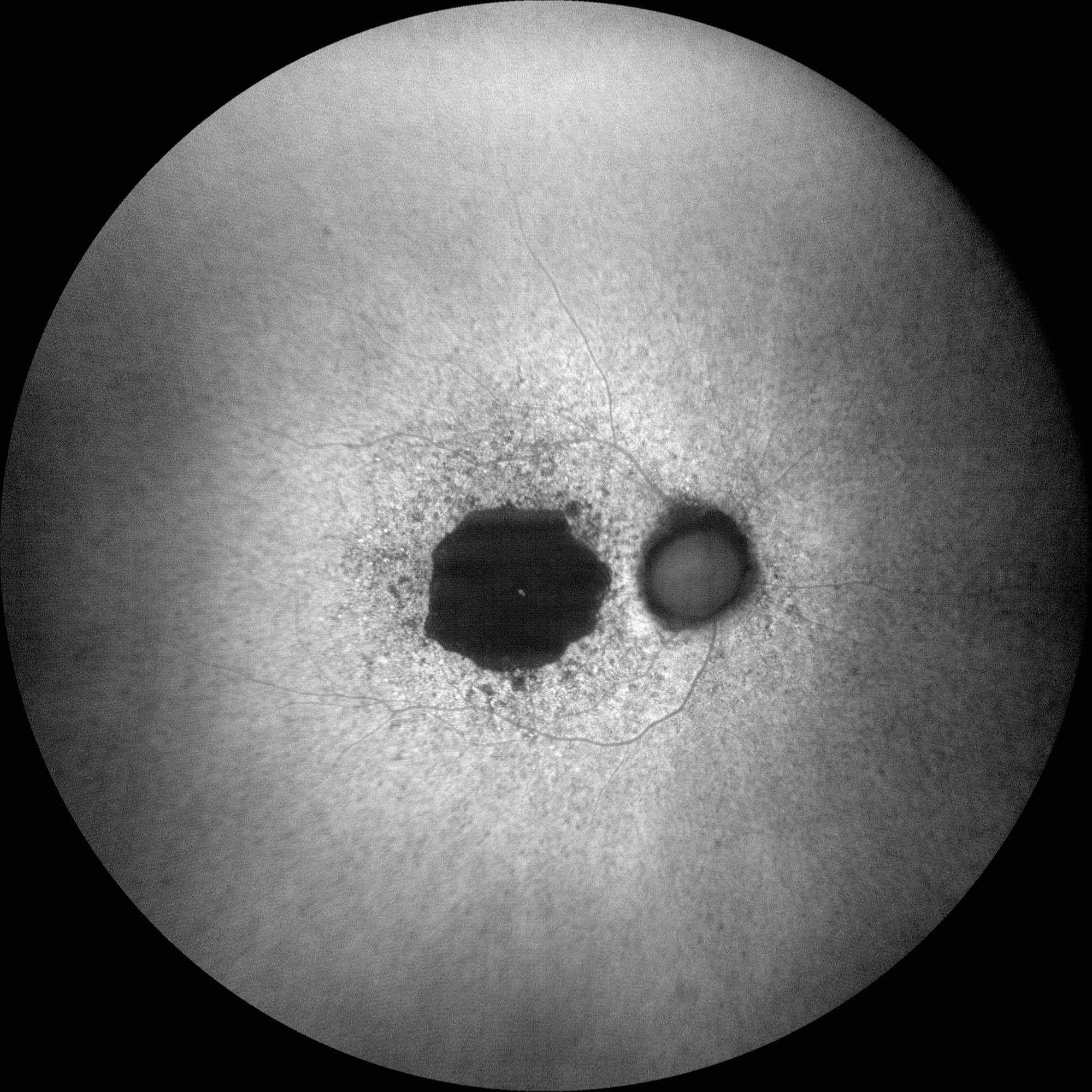
Green autofluorescence (Clarus 700, Zeiss): Hypoautofluorescent lesion corresponding to RPE atrophy. There is a mottled perilesional pattern suggesting metabolic stress of the RPE around the atrophy. A central hyperautofluorescent spot is also seen corresponding to the central healthy retinal island (B1, B2).

Green autofluorescence (Clarus 700, Zeiss): Hypoautofluorescent lesion corresponding to RPE atrophy. There is a mottled perilesional pattern suggesting metabolic stress of the RPE around the atrophy. A central hyperautofluorescent spot is also seen corresponding to the central healthy retinal island (B1, B2).
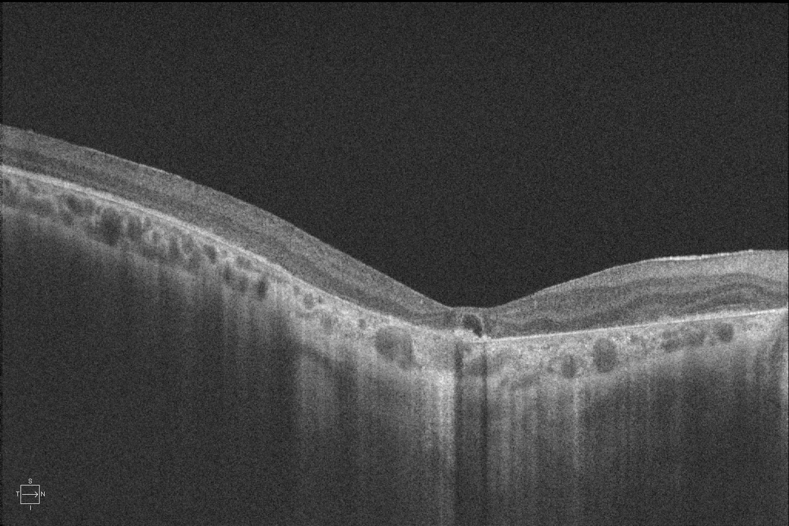
OCT (Cirrus 5000-HD, Zeiss): atrophy of the outer retina, RPE and choriocapillaris. What appears to be a foveal cyst in the right eye and a juxtafoveal cyst in the left eye is actually healthy retina (the hyporeflective part of this “pseudocyst” would be the layer of Henle fibers corresponding to the only area of preserved healthy retina) (C1, C2).
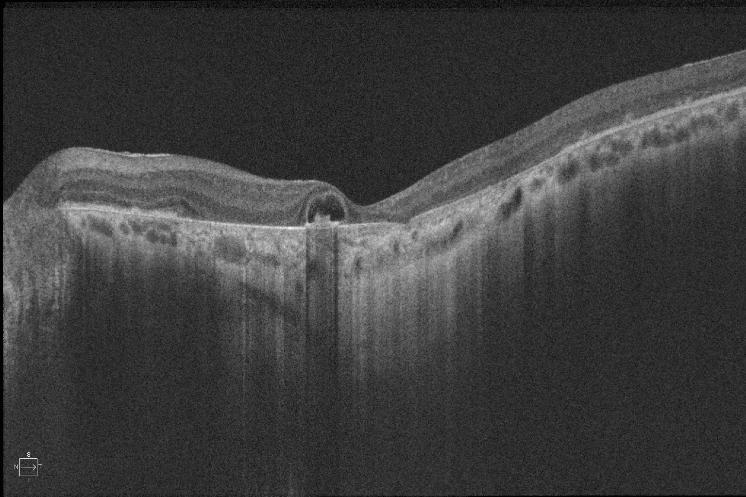
OCT (Cirrus 5000-HD, Zeiss): atrophy of the outer retina, RPE and choriocapillaris. What appears to be a foveal cyst in the right eye and a juxtafoveal cyst in the left eye is actually healthy retina (the hyporeflective part of this “pseudocyst” would be the layer of Henle fibers corresponding to the only area of preserved healthy retina) (C1, C2).
Description
A 45-year-old woman comes in for a check-up.
VA OD 20/400 OS 20/200.
In the fundus, central retinal atrophy with pigment deposition and visualization of underlying choroidal vessels is observed. The atrophy is hypoautofluorescent, with a mottled perilesional pattern and a central hyperautofluorescent spot. OCT shows complete atrophy of the outer retina, RPE, and choriocapillaris with a central island of healthy retina. The suspected diagnosis is Central Areolar Choroidal Dystrophy (CACD). The patient refused to undergo genetic testing.