Achromatopsia
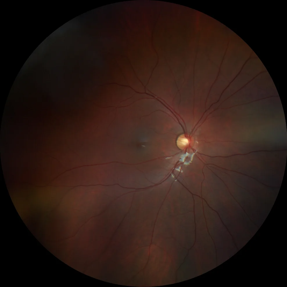
Achromatopsia A and B. Color retinography (Clarus 500, Carl Zeiss Meditec ASG, Jena, Germany) of the right and left eyes showing an anodyne fundus, in which only an isolated congenital hyperplasia of the retinal pigment epithelium in the inferotemporal area of the left eye stands out
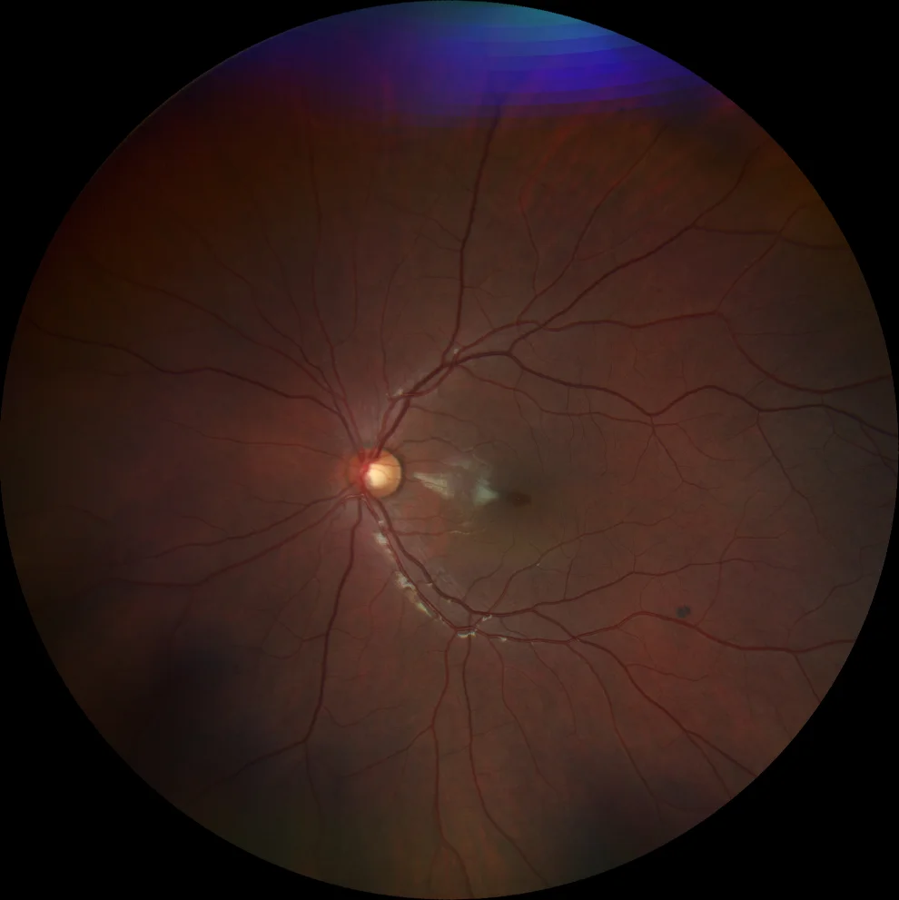
Achromatopsia A and B. Color retinography (Clarus 500, Carl Zeiss Meditec ASG, Jena, Germany) of the right and left eyes showing an anodyne fundus, in which only an isolated congenital hyperplasia of the retinal pigment epithelium in the inferotemporal area of the left eye stands out
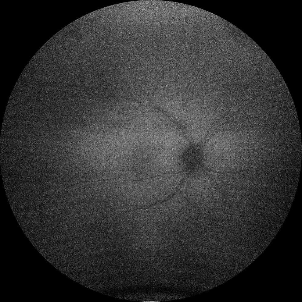
C and D. Autofluorescence (Clarus 500, Carl Zeiss Meditec ASG, Jena, Germany) of the right and left eyes also without relevant findings.
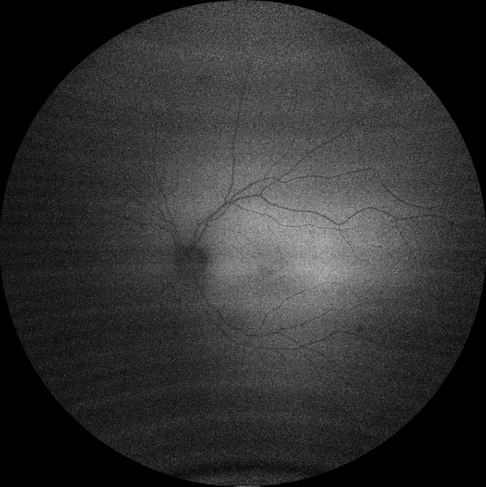
C and D. Autofluorescence (Clarus 500, Carl Zeiss Meditec ASG, Jena, Germany) of the right and left eyes also without relevant findings.
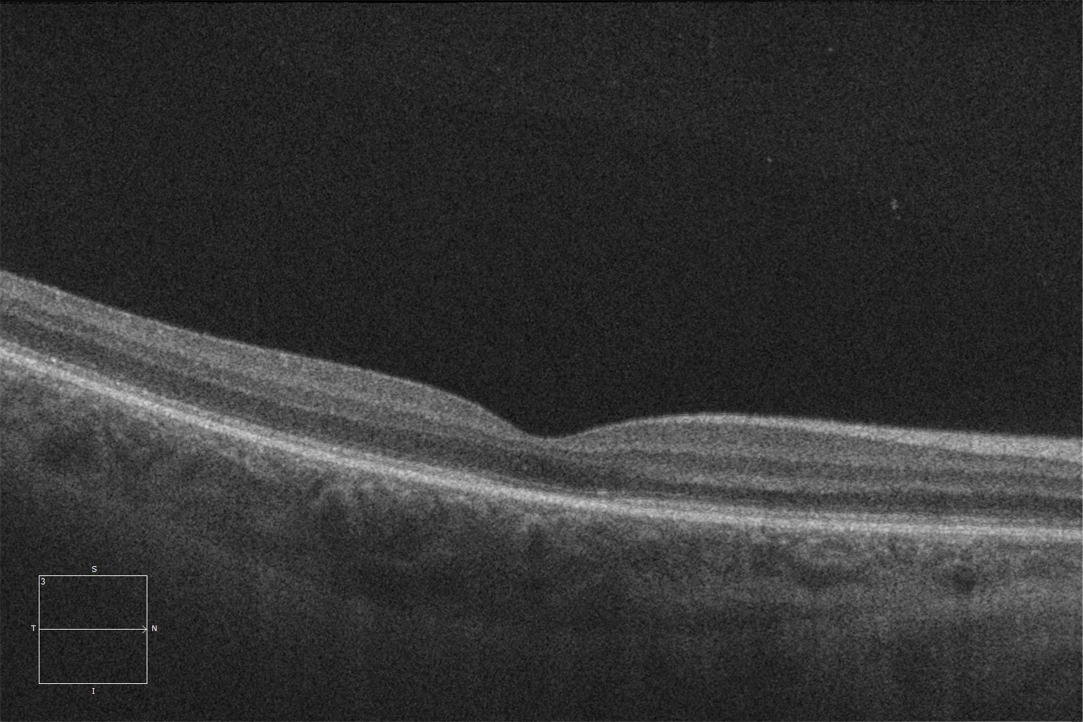
E and F. Optical coherence tomography (Cirrus 5000, Carl Zeiss Meditec ASG, Jena, Germany) of the macular area in the right and left eyes showing attenuation of the ellipsoid layer in the subfoveal area.
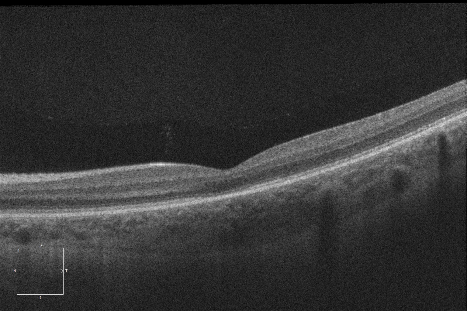
E and F. Optical coherence tomography (Cirrus 5000, Carl Zeiss Meditec ASG, Jena, Germany) of the macular area in the right and left eyes showing attenuation of the ellipsoid layer in the subfoveal area.
Description
Achromatopsia is a stationary congenital defect characterized by selective dysfunction of the cone system. It is a rare congenital disease of genetic origin with autosomal recessive inheritance, having an incidence of 1/30,000 to 1/50,000. 90% of affected patients present mutations in the CNGA3 or CNGB3 genes, which encode the CNG channel located exclusively in the plasma membrane of cones and is fundamental in the phototransduction cascade. The classic presentation includes early low visual acuity, profound achromatopsia, congenital nystagmus, and photophobia. The fundus examination is usually unremarkable. In addition to genetic testing, electrophysiological study is essential in the diagnosis of these patients. The full-field electroretinogram (ffERG) shows a severe decrease or abolition of the photopic response and 30 Hz flicker, with a characteristically preserved scotopic signal. High-definition OCT may reveal defects in the subfoveal ellipsoid zone. Although no treatment has yet been approved for this disease, there are several clinical trials based on genetic editing with recombinant adeno-associated viruses that show promising preliminary results. For now, the management of these patients is limited to genetic counseling, low vision aids, early specific treatment of amblyopia and nystagmus, and filters to reduce photophobia.