Ocular ischemic syndrome
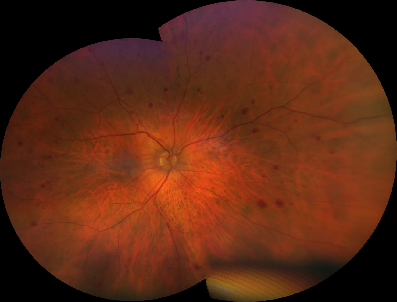
Retinography (Clarus 700, Zeiss): deep peripheral (blot) hemorrhages and microaneurysms in 360º of both eyes
Retinography (Clarus 700, Zeiss): deep peripheral (blot) hemorrhages and microaneurysms in 360º of both eyes
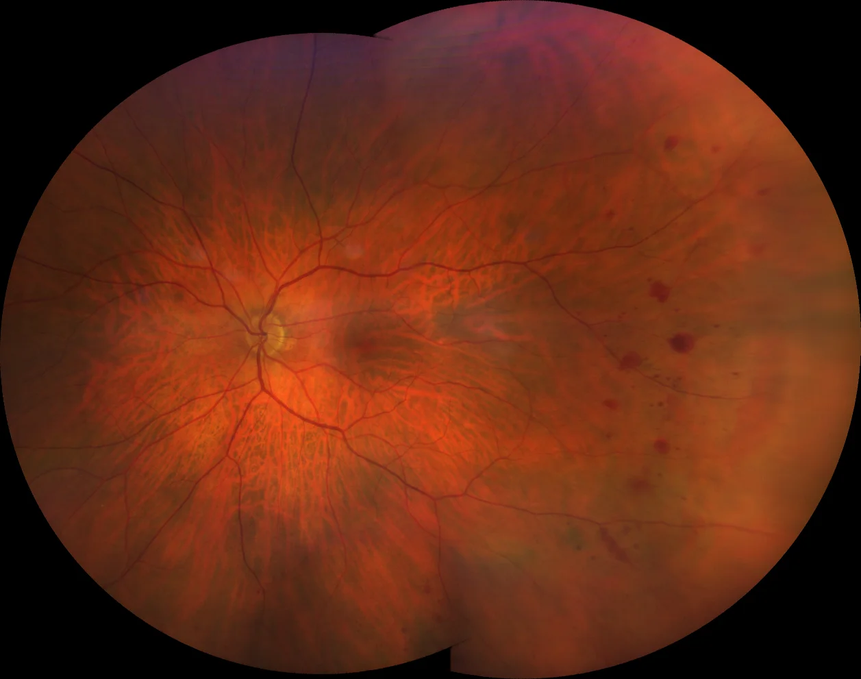
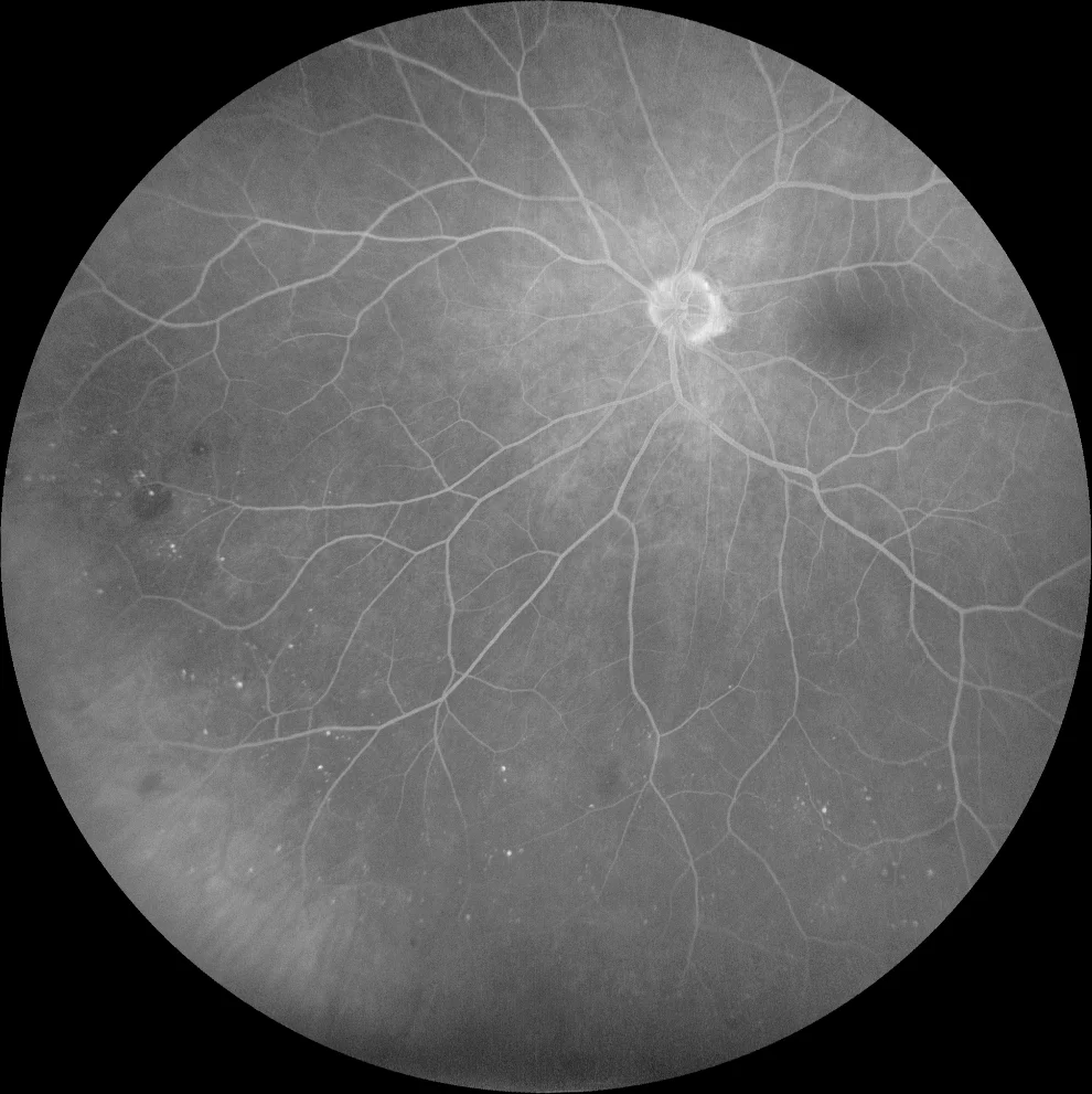
Four months after surgery, the hemorrhages have disappeared, although some microaneurysms are still observed
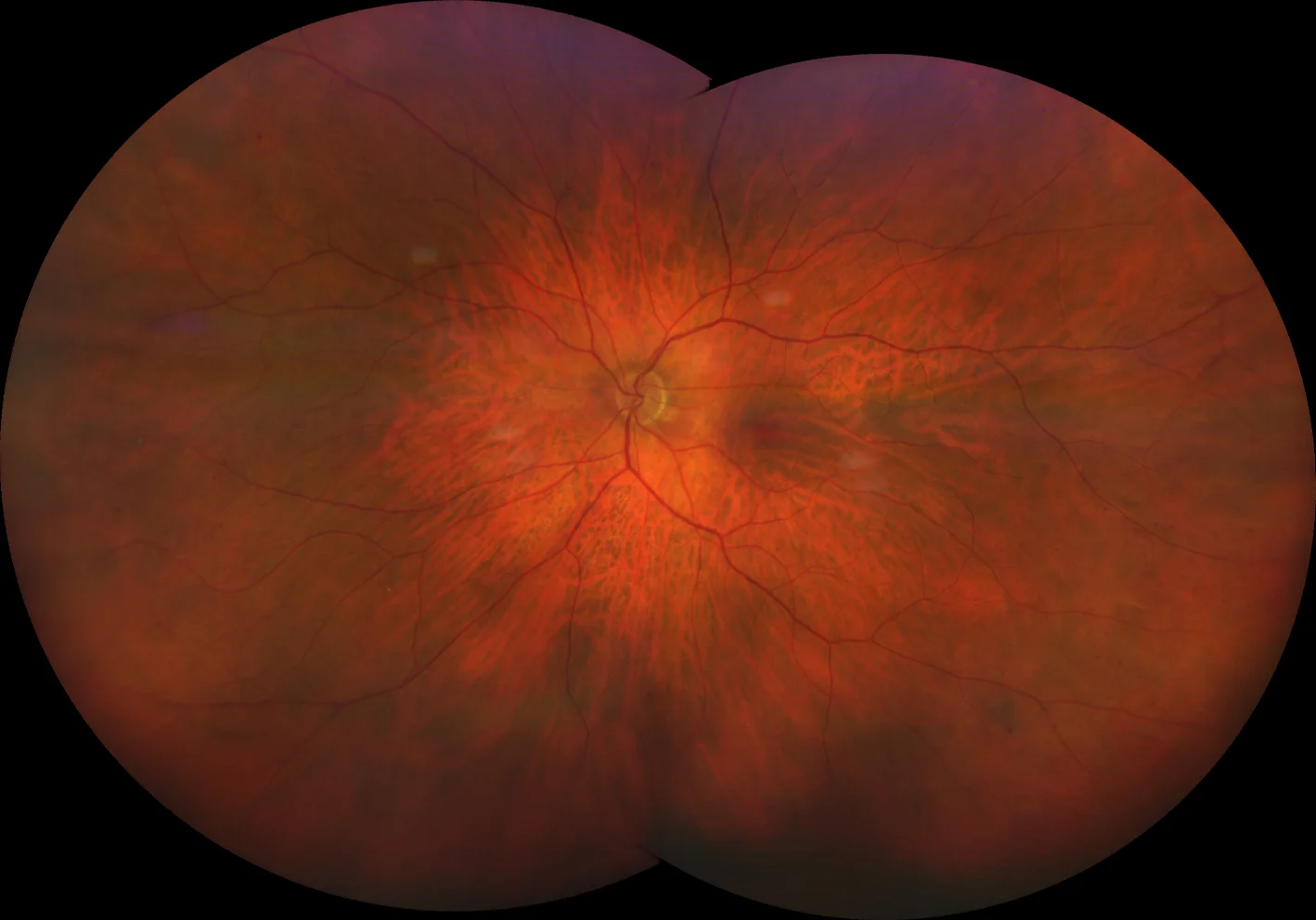
Four months after surgery, the hemorrhages have disappeared, although some microaneurysms are still observed
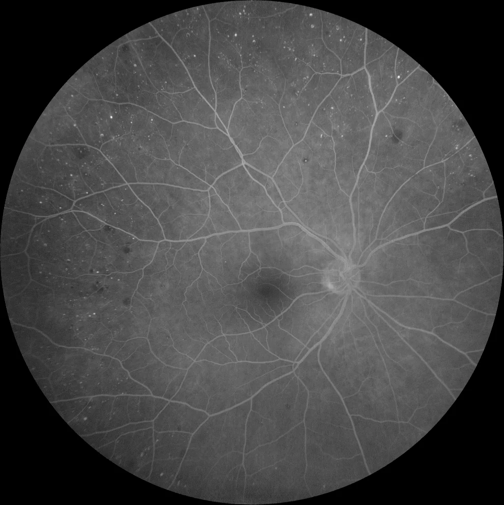
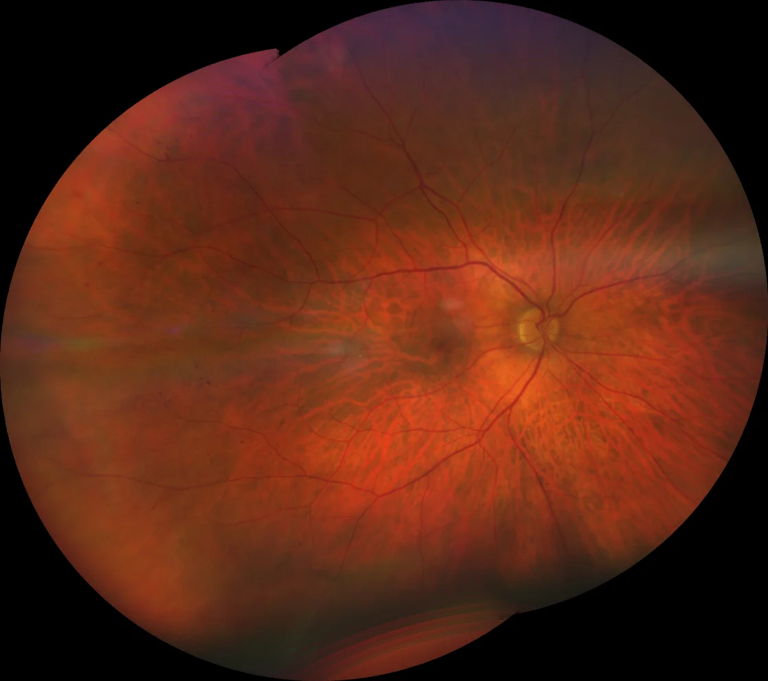
FAG (Clarus 700, Zeiss): numerous peripheral microaneurysms in both eyes
FAG (Clarus 700, Zeiss): numerous peripheral microaneurysms in both eyes
Description
An 80-year-old male presents with floaters in both eyes.
Visual acuity is 20/25 in both eyes. Examination of the anterior segment is unremarkable. Fundus examination reveals multiple dot and blot hemorrhages and what appear to be microaneurysms in the 360° periphery of both eyes. Fluorescein angiography confirms the presence of numerous peripheral microaneurysms in 360° in both eyes. No macular edema is observed on OCT. In addition to ruling out DM, hypertension, and blood dyscrasias, the patient is referred to a vascular surgeon who, on Doppler ultrasound of the supra-aortic trunks, finds severe stenosis in both internal carotid arteries. The diagnosis of bilateral ocular ischemic syndrome is confirmed. Four months after endarterectomy surgery, the hemorrhages have disappeared, with some microaneurysms persisting.