Adult-Onset Foveomacular Vitelliform Dystrophy
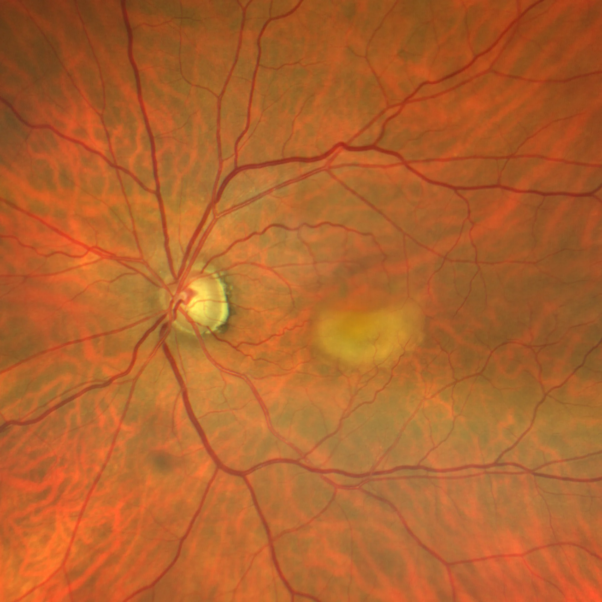
Color (detail): Yellowish rounded macular lesion
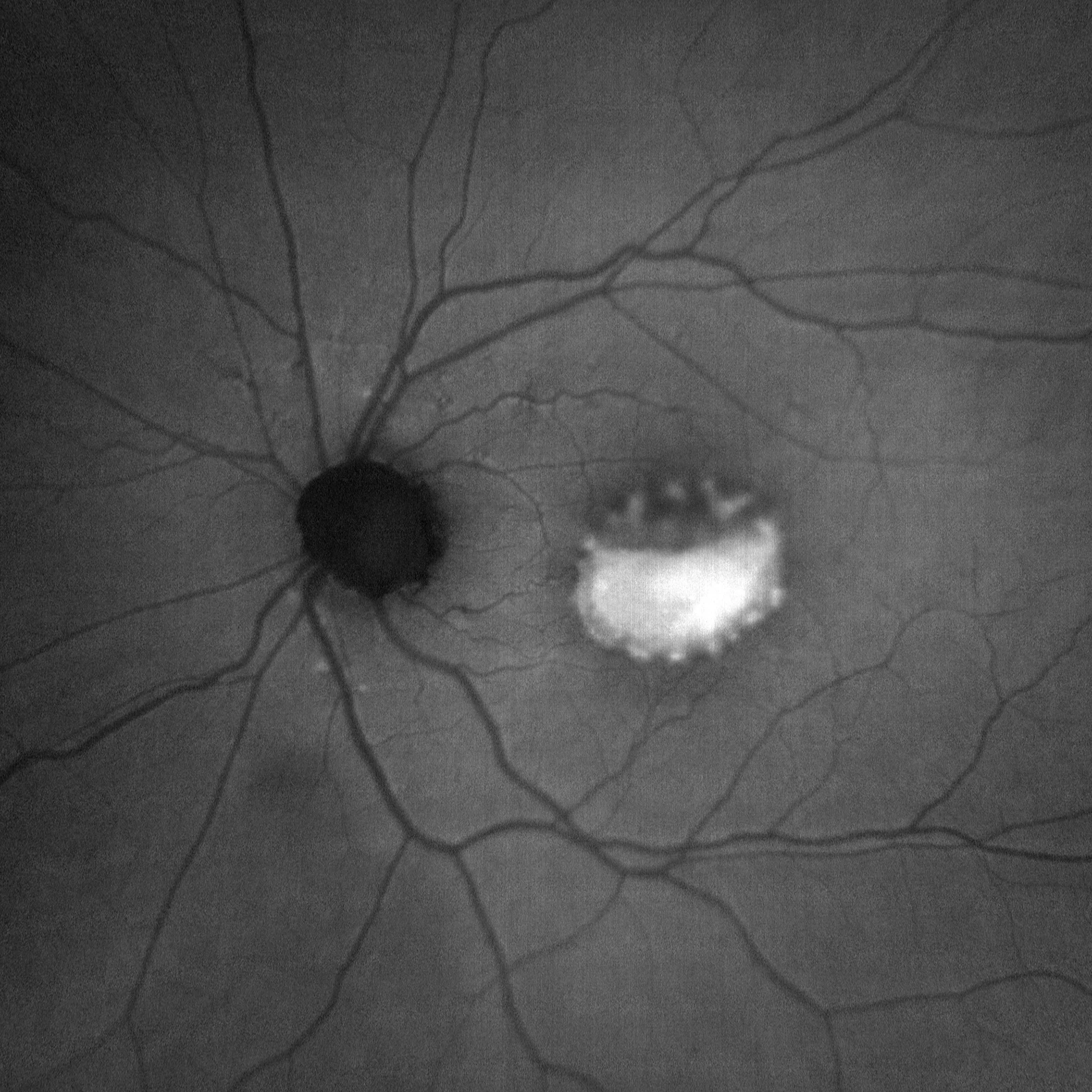
Green autofluorescence (detail): Marked autofluorescence of vitelliform material
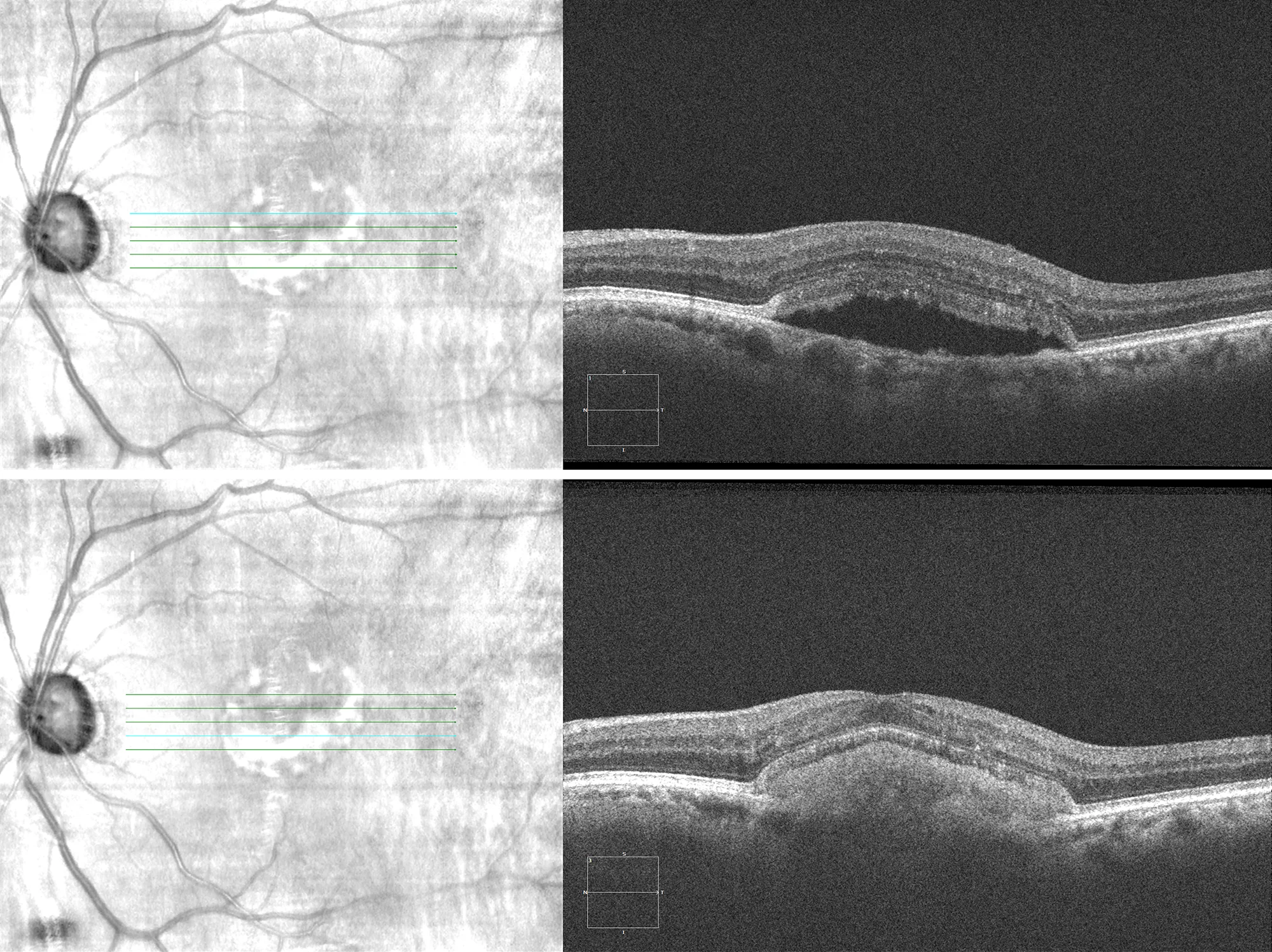
Optical coherence tomography (Cirrus-HD 5000): Rounded lesion between retinal pigment epithelium and neurosensory retina with vitelliform material in the inferior zone.
Description
Adult-onset foveomacular vitelliform dystrophy is characterized by bilateral, rounded, yellowish lesions at the macular level, with an appearance similar to Best’s disease. Patients are typically diagnosed in adulthood due to a slow and progressive loss of vision. Autofluorescence and OCT findings are very characteristic and allow differentiation from other forms of macular degeneration. The disease progresses through various stages that ultimately lead to macular atrophy.