< back
Epiretinal Membrane (ERM)
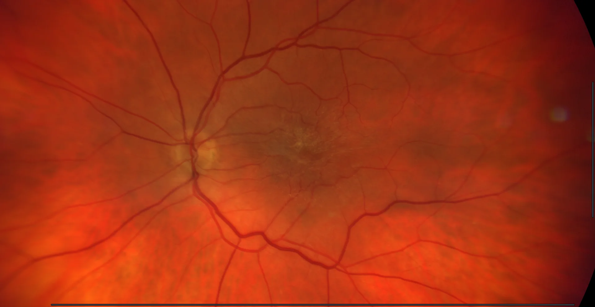
Color retinography (CLARUS 500, Zeiss): Epiretinal membrane at the macular level with retinal folds.
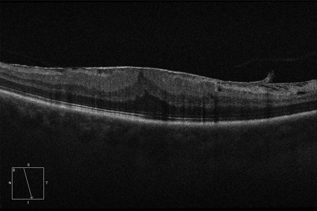
OCT (Cirrus-HD 6000, Zeiss): Macular epiretinal membrane altering the foveal profile. The posterior hyaloid is partially detached.
Description
Epiretinal Membrane (ERM). It is an avascular, transparent fibrocellular tissue located on the internal surface of the retina that adheres to and covers the internal limiting membrane. It is usually located at the macular level and can be bilateral in up to 20% of cases. In most cases, it is associated with the presence of posterior vitreous detachment, as it is believed to be a key factor in the pathophysiology of the condition.