Best Disease
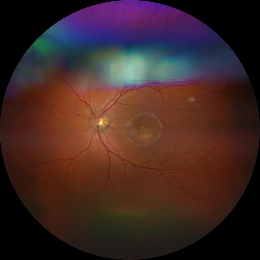
Retinography (Clarus 700, Zeiss): macular atrophy with inferior vitelliform deposits (A) is observed in the left eye. The right eye could not be recorded due to lack of cooperation of the patient due to intense photophobia.
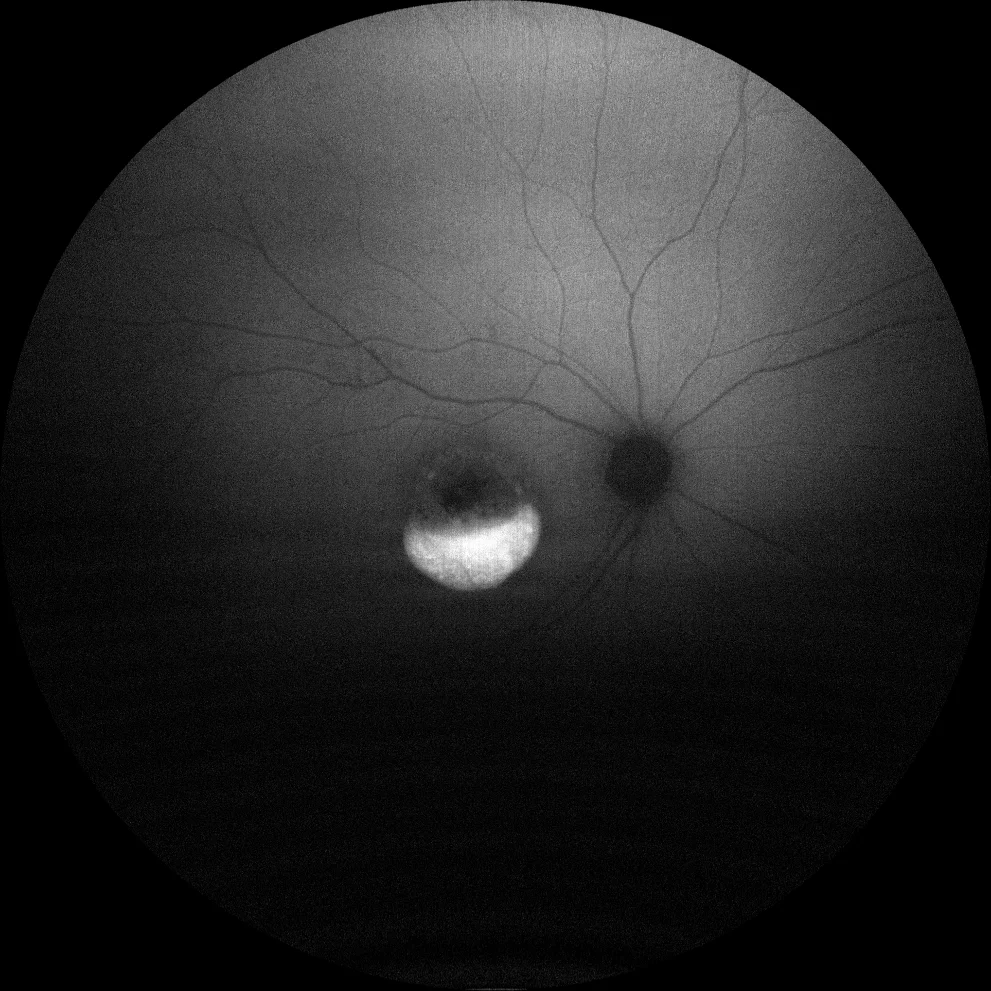
Green autofluorescence (Clarus 700, Zeiss): central RPE atrophy with hyperautofluorescent vitelliform material at the lower level (B1, B2).
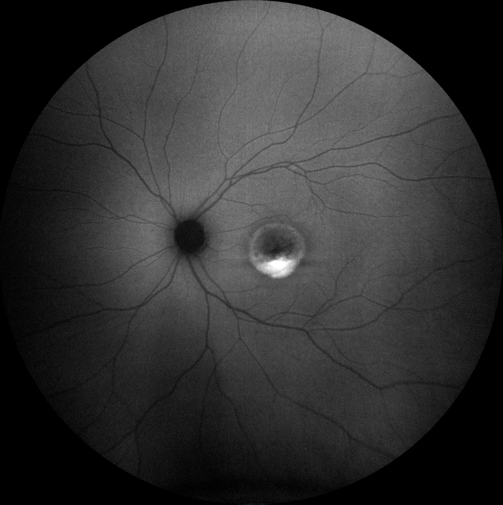
Green autofluorescence (Clarus 700, Zeiss): central RPE atrophy with hyperautofluorescent vitelliform material at the lower level (B1, B2).
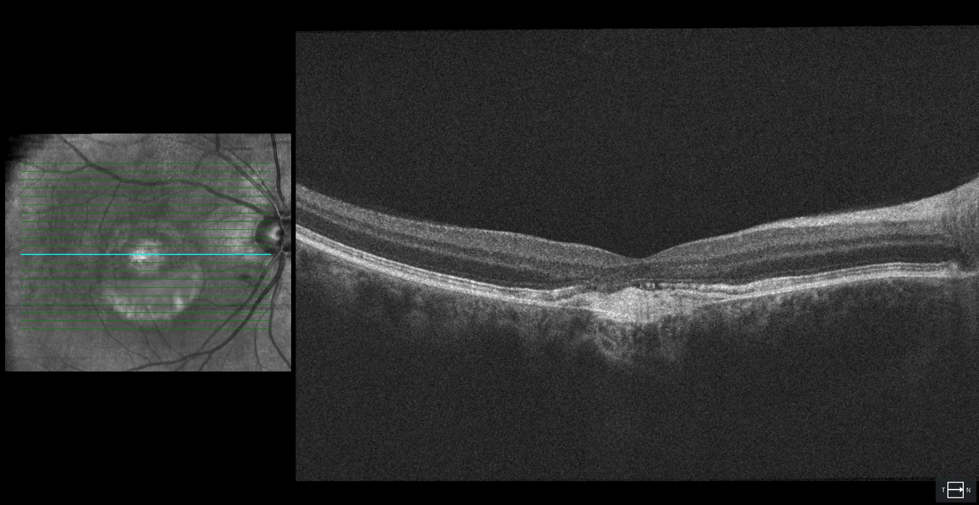
OCT (Cirrus 5000-HD, Zeiss) and OCT-angiography (Cirrus 5000-HD, Angioplex, Zeiss): in the right eye, a disruption of the external limiting membrane, ellipsoid zone (EZ) and interdigitation zone (IZ) at the central level (C1) is observed.
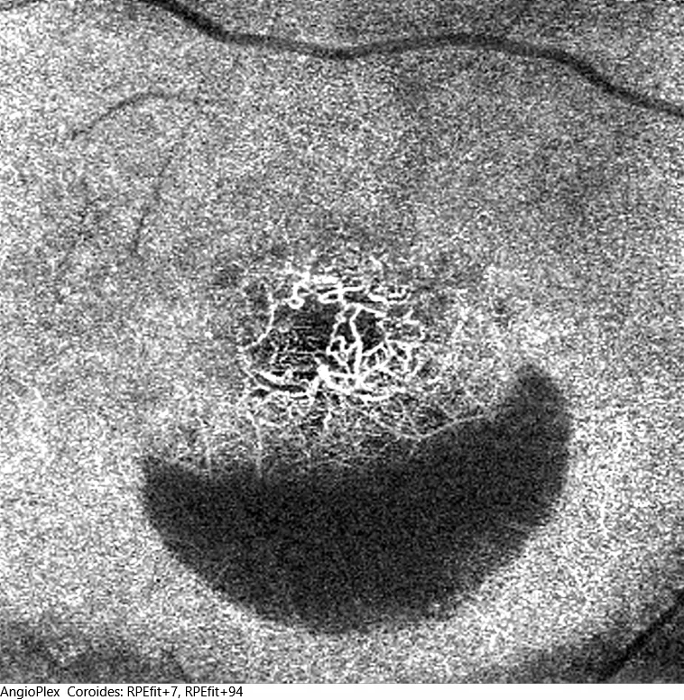
Hyperreflective material is also seen beneath the RPE with a break in Bruch's membrane. This suggests the presence of choroidal neovascularization, which is confirmed by OCT-A (C2).
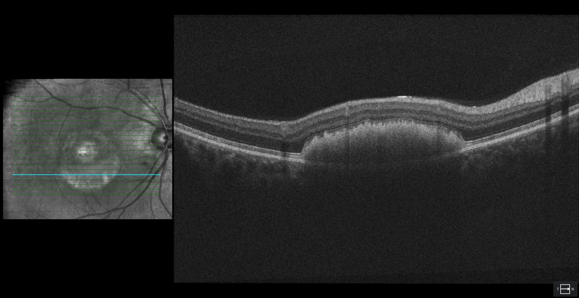
A lower section in the same eye shows a subretinal hyperreflective material corresponding to the vitelliform material (C3).
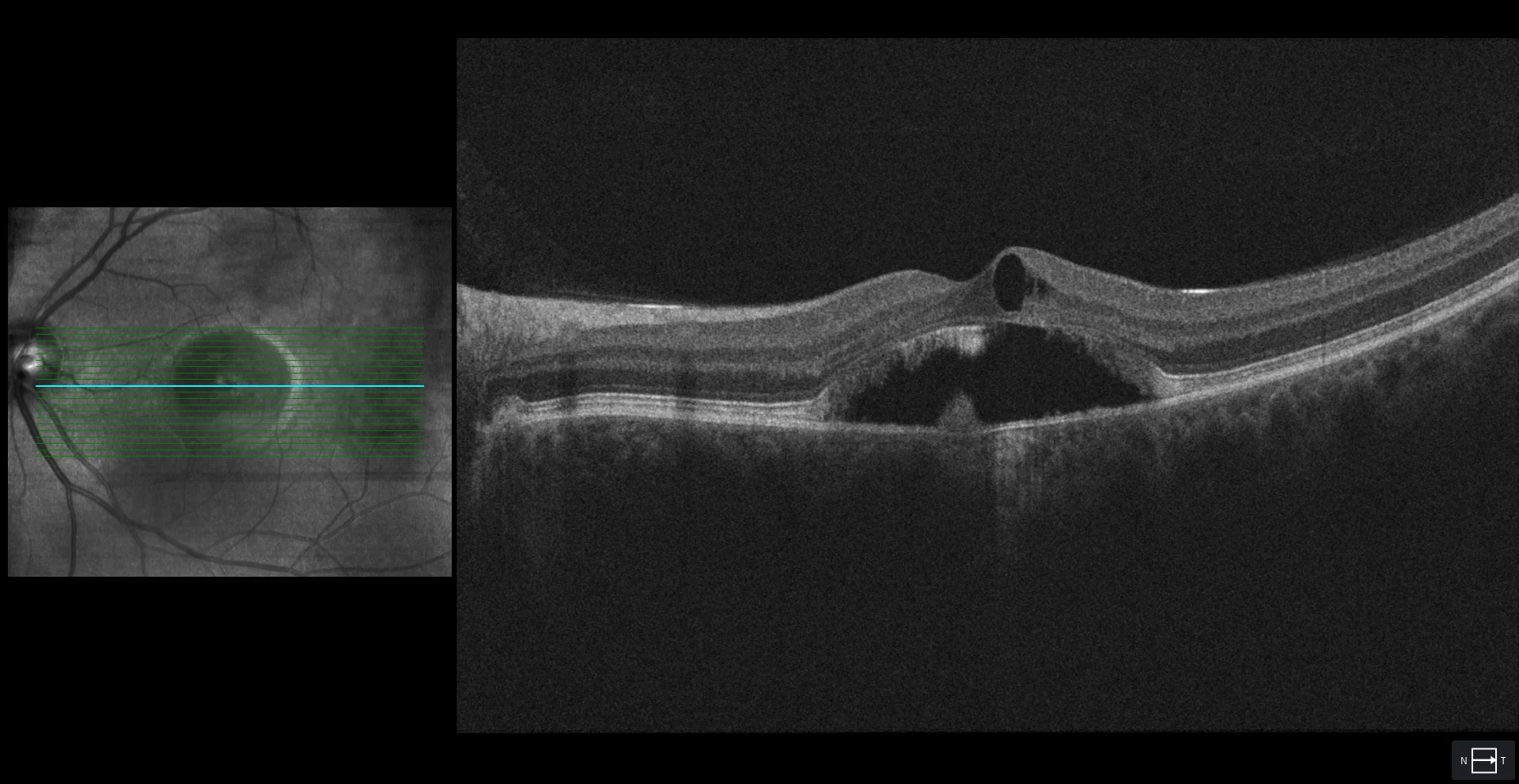
In the left eye, the foveal section shows subretinal fluid, as well as a minimal amount of intraretinal fluid; elongation of the photoreceptors (brush border pattern) and areas of disruption of the EZ and IZ (C4) are observed.
Description
A 38-year-old male presents with progressive vision loss over the past few months.
VA OD 20/25 OS 20/25.
Fundus examination reveals a bilateral macular lesion with a yellowish inferior deposit (with a level) in both eyes. This deposit is hyperautofluorescent. The OCT suggests the presence of macular neovascularization in the right eye, which is confirmed by OCT-A. The suspected diagnosis is Best disease, so the patient is advised to undergo genetic testing.