Stargardt Disease
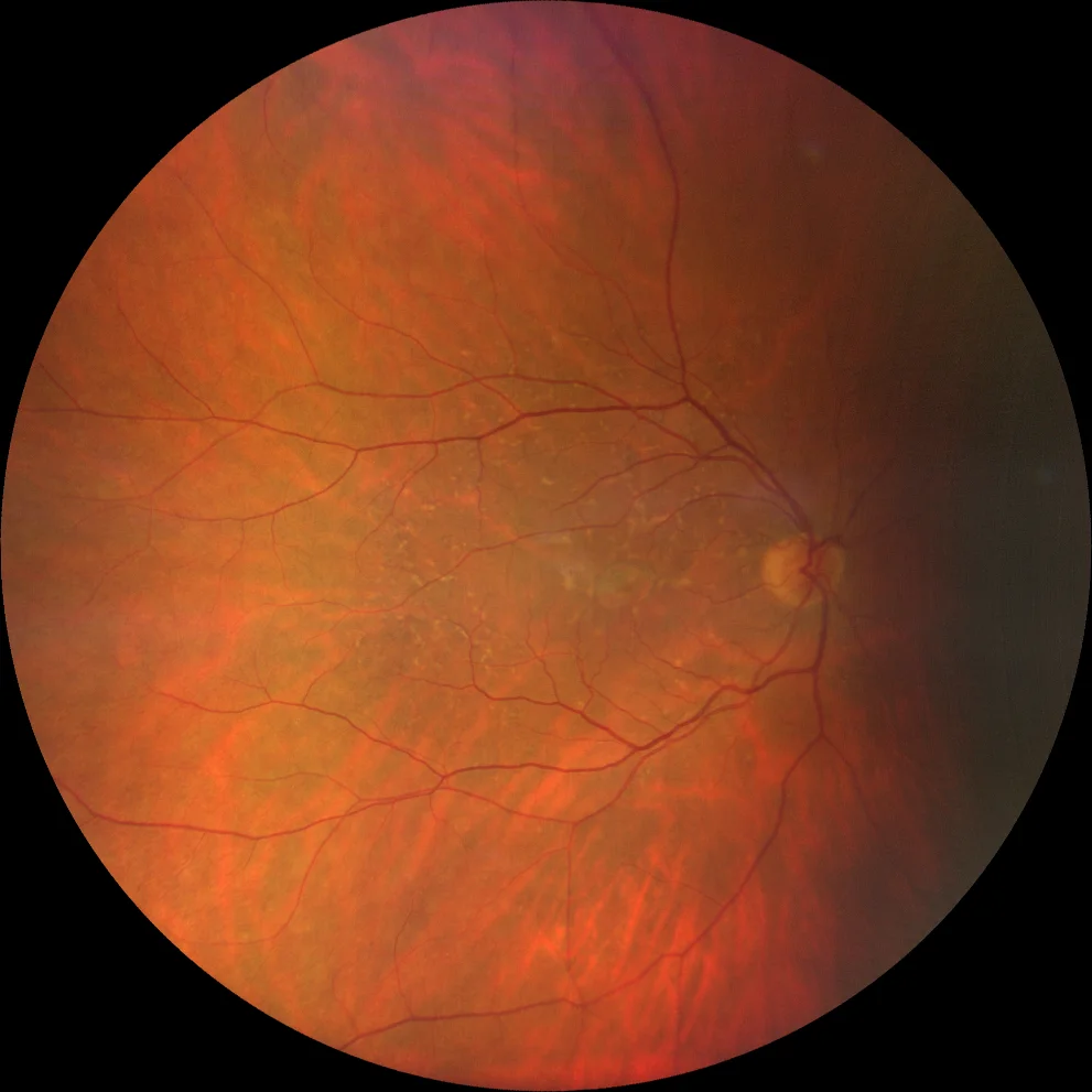
Retinography (Clarus 700, Zeiss): pisciform flecks in the macula and nasal area to the papilla in both eyes (A1, A2). Central atrophy is also observed in the right eye (A1).
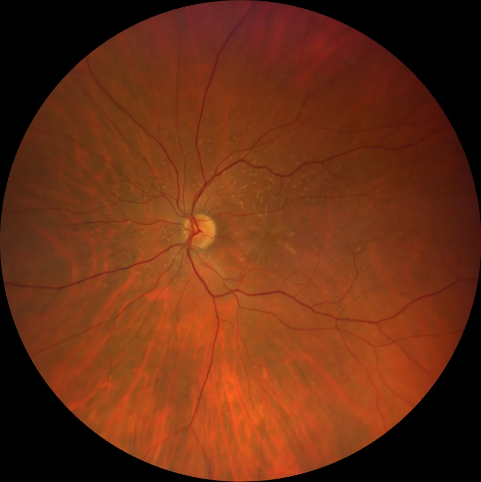
Retinography (Clarus 700, Zeiss): pisciform flecks in the macula and nasal area to the papilla in both eyes (A1, A2). Central atrophy is also observed in the right eye (A1).
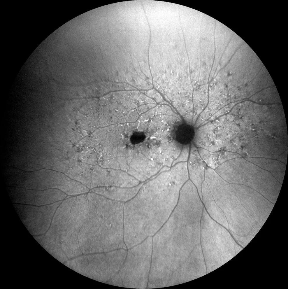
Green autofluorescence (Clarus 700, Zeiss): Flecks are hyperautofluorescent due to their high lipofuscin content, although there are also hypofluorescent lesions due to RPE atrophy (B1, B2). Central atrophy is more evident in AF than in GR (B1).
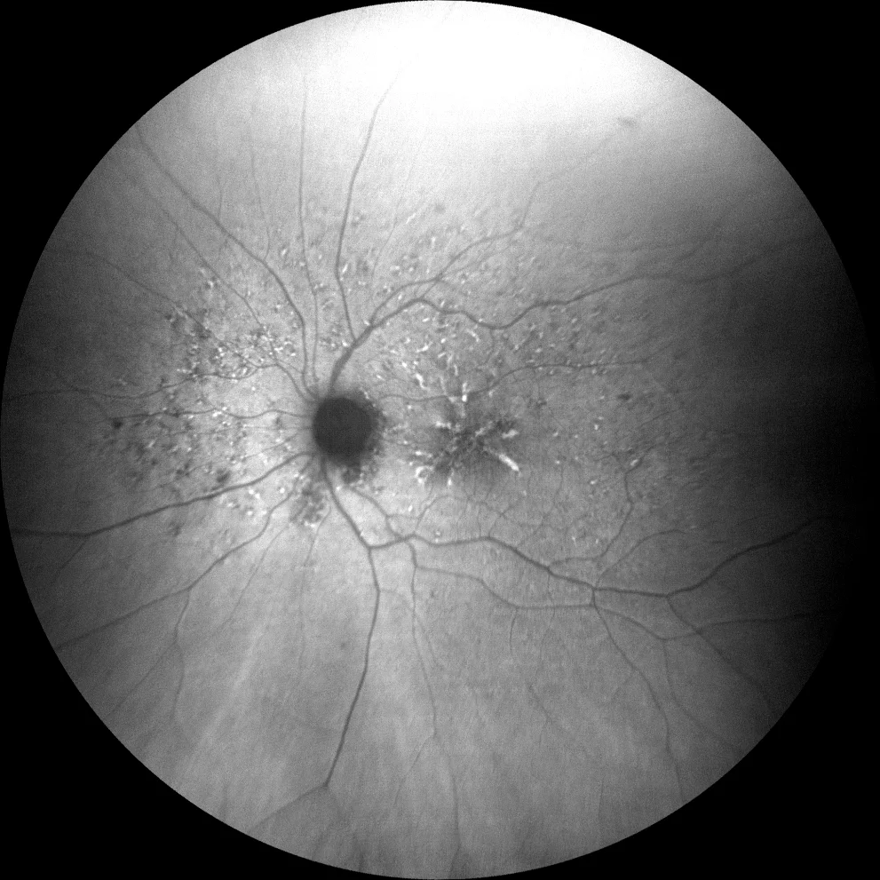
Green autofluorescence (Clarus 700, Zeiss): Flecks are hyperautofluorescent due to their high lipofuscin content, although there are also hypofluorescent lesions due to RPE atrophy (B1, B2). Central atrophy is more evident in AF than in GR (B1).
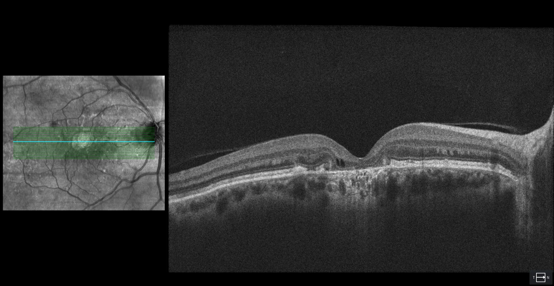
OCT (Cirrus 5000-HD, Zeiss): atrophy of the outer retina at the foveal level in the right eye (C1). In the left eye, the outer retina is preserved, which explains the patient's good visual acuity in that eye (C2). In this case, the thickened MLE, typical of Stargardt disease with onset in childhood, is not observed.
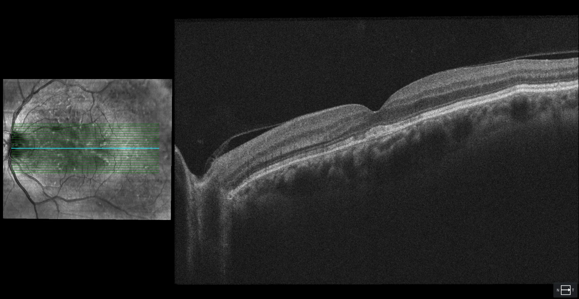
OCT (Cirrus 5000-HD, Zeiss): atrophy of the outer retina at the foveal level in the right eye (C1). In the left eye, the outer retina is preserved, which explains the patient's good visual acuity in that eye (C2). In this case, the thickened MLE, typical of Stargardt disease with onset in childhood, is not observed.
Description
67-year-old woman who comes for a check-up.
VA OD 20/100 OS 20/20.
In the fundus of both eyes, yellowish deposits (flecks) are observed in the posterior pole and in the area nasal to the optic disc. These deposits are more evident in autofluorescence, which also reveals foveal atrophy in the right eye. OCT shows atrophy of the outer retina at the foveal level in the right eye and a hyperreflective subfoveal deposit with preservation of the ELM and EZ in the left eye.