X-linked juvenile retinoschisis
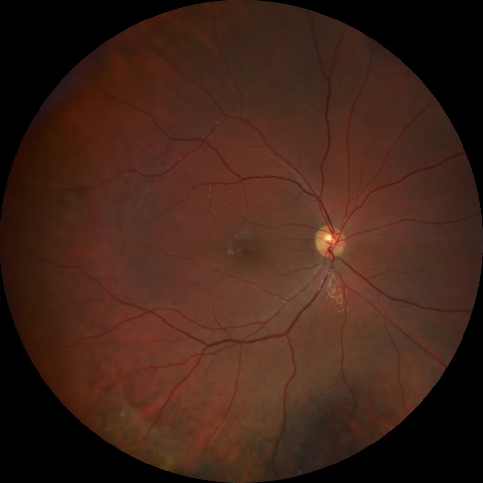
A. Color retinography (Clarus 500, Carl Zeiss Meditec ASG, Jena, Germany) of the RE showing macular radial striae.
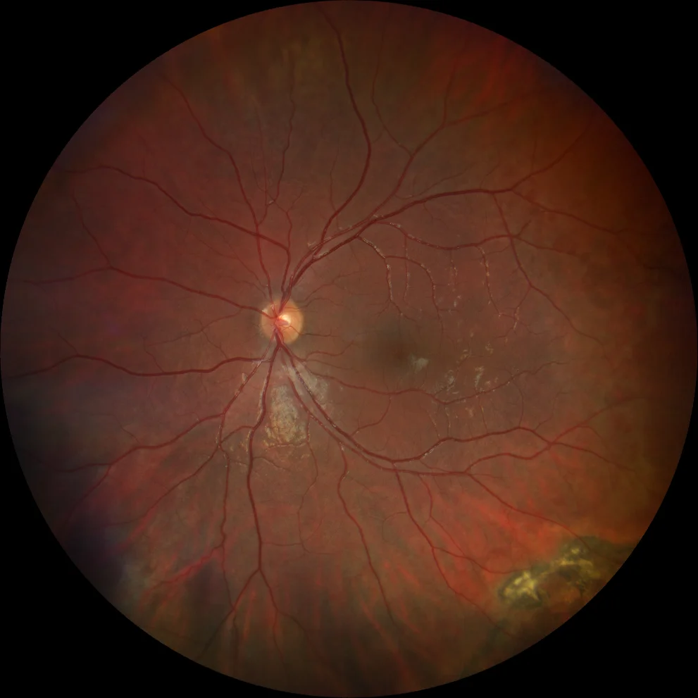
B. Color retinography (Clarus 500, Carl Zeiss Meditec ASG, Jena, Germany) of the LE in which pigmentary changes and subretinal fibrosis are evident in the infero-temporal quadrant
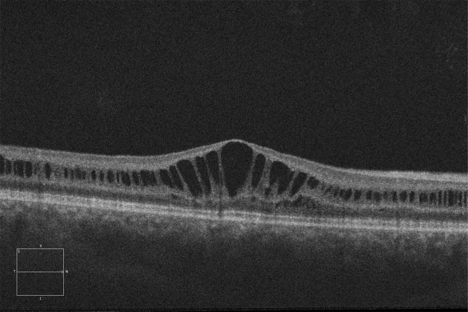
C and D. Macular HD optical coherence tomography (Cirrus 5000, Carl Zeiss Meditec ASG, Jena, Germany) of the right and left eyes, showing multiple macular cystic spaces located at the level of the inner nuclear layer.
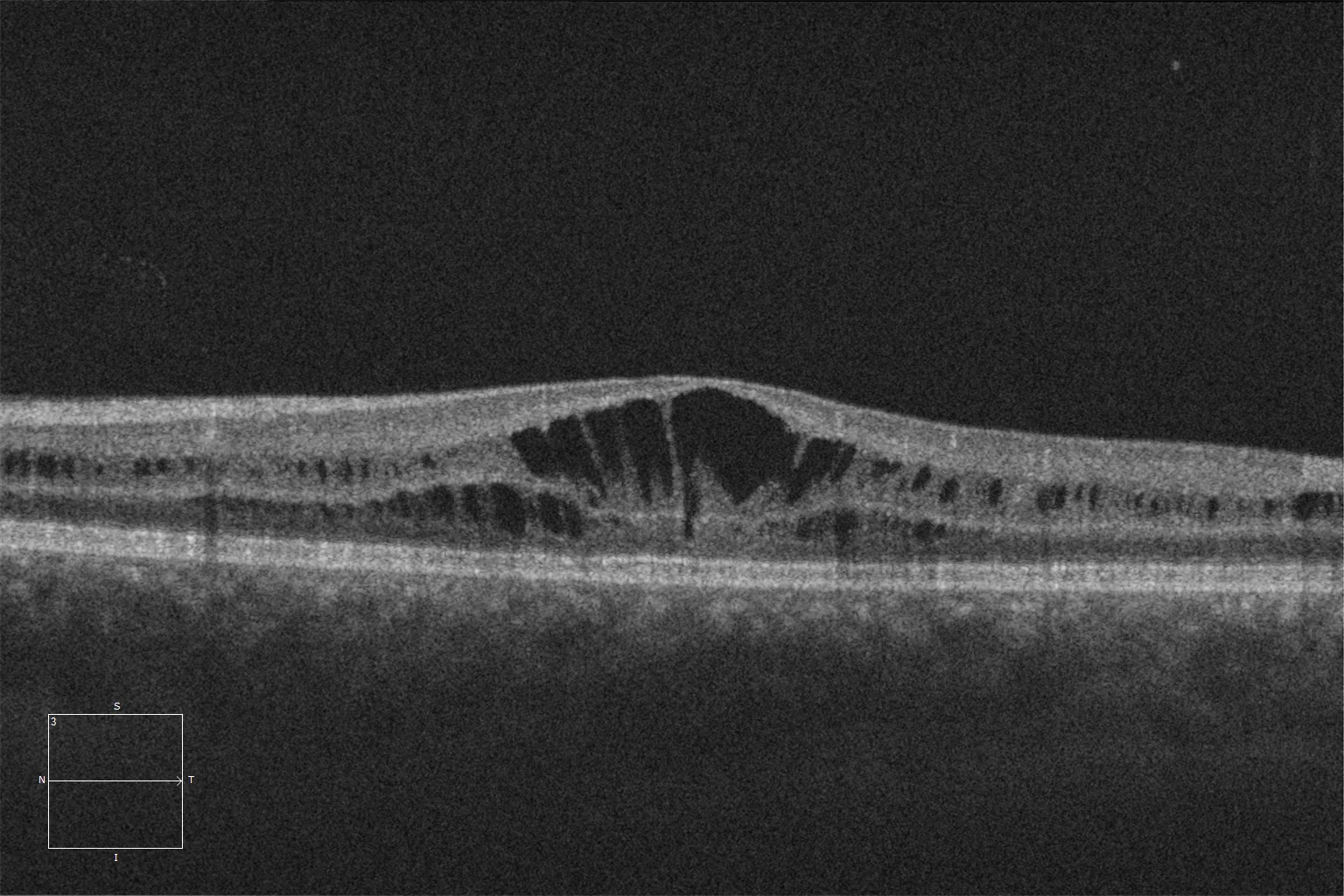
C and D. Macular HD optical coherence tomography (Cirrus 5000, Carl Zeiss Meditec ASG, Jena, Germany) of the right and left eyes, showing multiple macular cystic spaces located at the level of the inner nuclear layer.
Description
X-linked retinoschisis (XLRS) is a vitreoretinal degeneration caused by various mutations in the RS1 gene. It predominantly affects males and usually presents in the first decade of life with vision loss, which often remains stable until the fifth or sixth decades. In infants, it can present with strabismus, nystagmus, or amblyopia. Bilateral foveal schisis is the most common finding. 50% of patients also present with peripheral schisis, typically located in the inferotemporal quadrant. Fine striations radiating from the fovea can be observed in the fundus. Pigmentary changes, vitreous veils, and peripheral dendritic lesions may also appear. Optical coherence tomography (OCT) can reveal macular cystic spaces. Fundus autofluorescence may show foveal hyperautofluorescence. Some patients may develop complications such as vitreous hemorrhage, neovascularization, or rhegmatogenous or tractional retinal detachment.