Multiple Evanescent White Dot Syndrome (MEWDS)
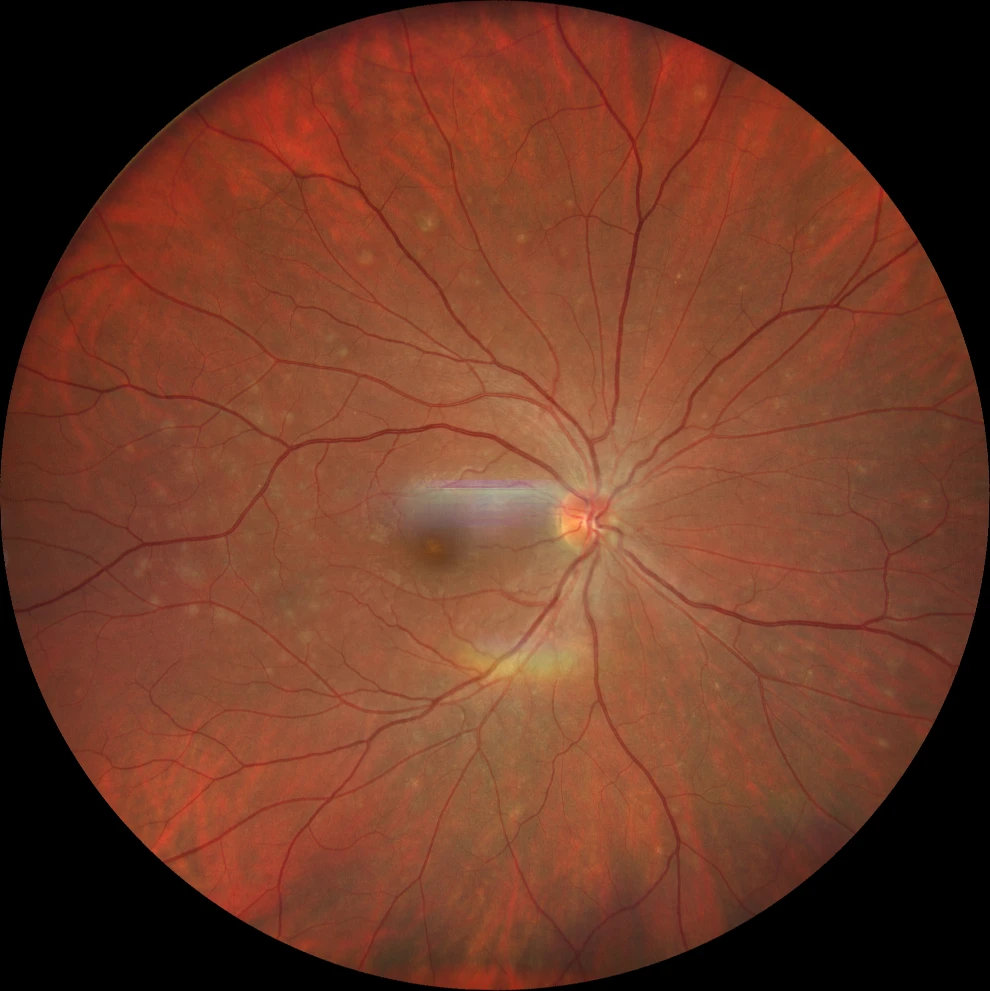
A. Color retinography (Clarus 500, Carl Zeiss Meditec ASG, Jena, Germany) showing multiple yellowish-white lesions reaching the middle periphery and a granular appearance in the fovea.
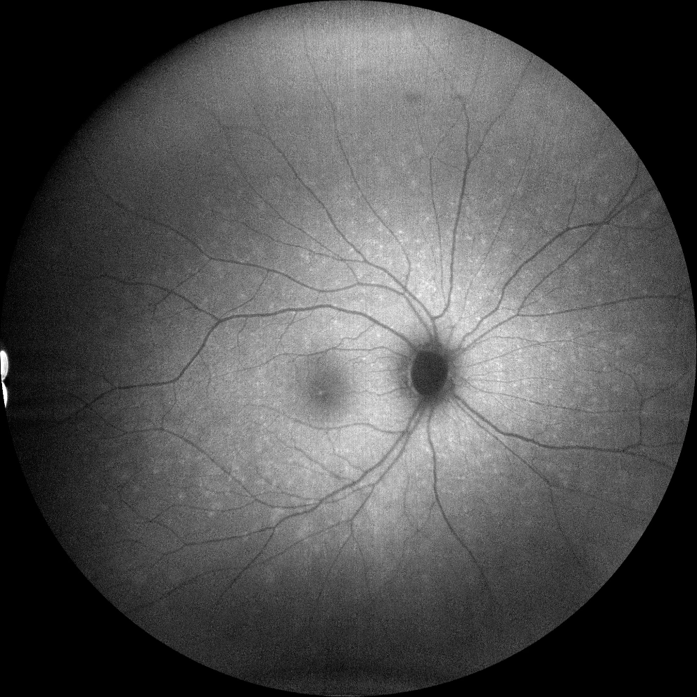
B. Autofluorescence image (Clarus 500, Carl Zeiss Meditec ASG, Jena, Germany) showing more hyperautofluorescent lesions than those present in the color retinography.
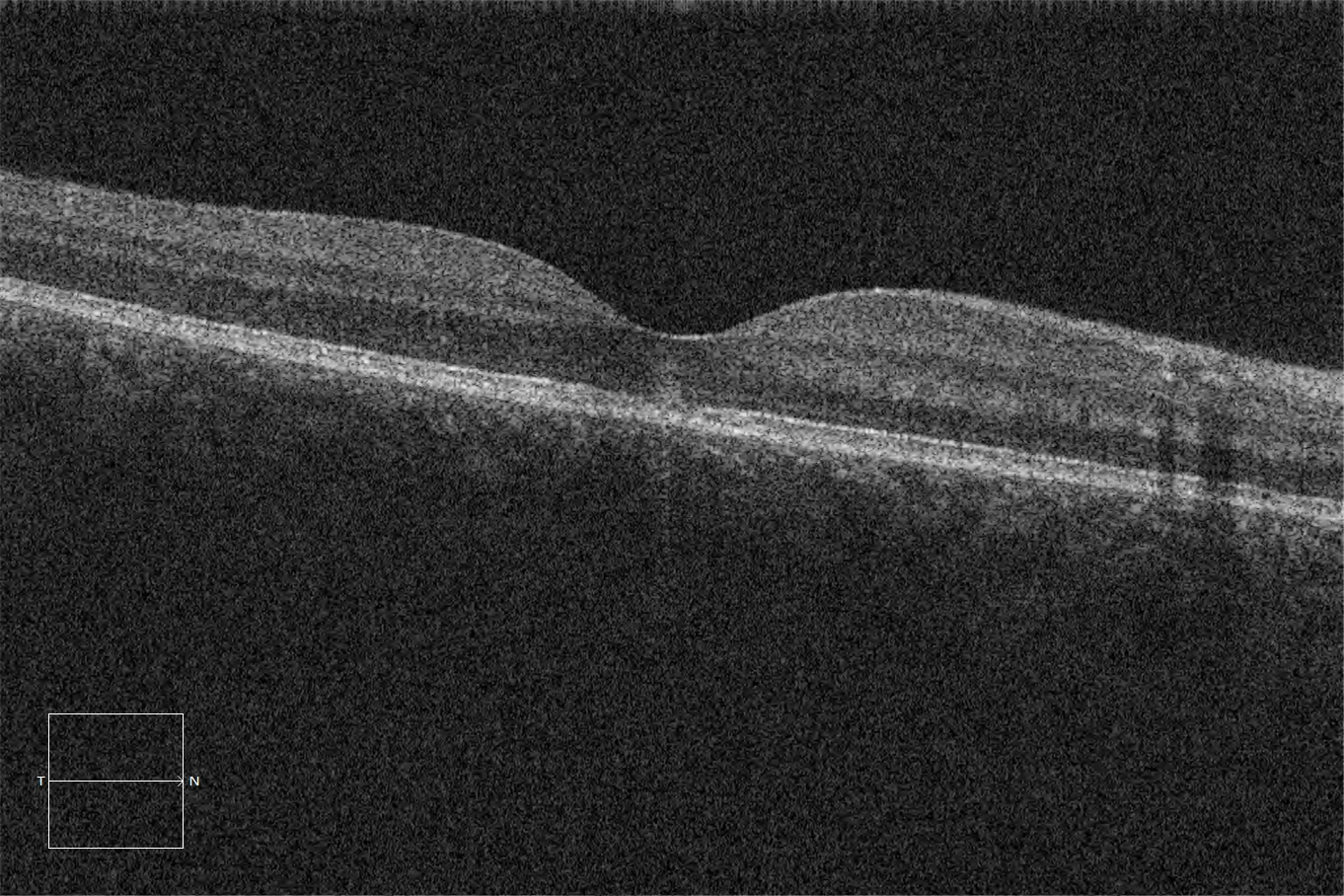
C. Macular optical coherence tomography (Cirrus 5000, Carl Zeiss Meditec ASG, Jena, Germany) with loss of the ellipsoid and subfoveal external limiting membrane due to the presence of a hyperrefringent image at that level.
Description
MEWDS is a rare entity of unknown etiology that typically affects young women. The most characteristic finding in the fundus is the presence of multiple white-yellowish lesions of 100-200 μm that reach the mid-periphery. The macula usually acquires a granular appearance that persists longer than the multiple evanescent whitish lesions. In autofluorescence, hyperautofluorescent lesions with poorly defined borders can be observed. In optical coherence tomography, the presence of hyperreflective lesions in outer layers is characteristic. The condition is usually unilateral.