Angioid Streaks
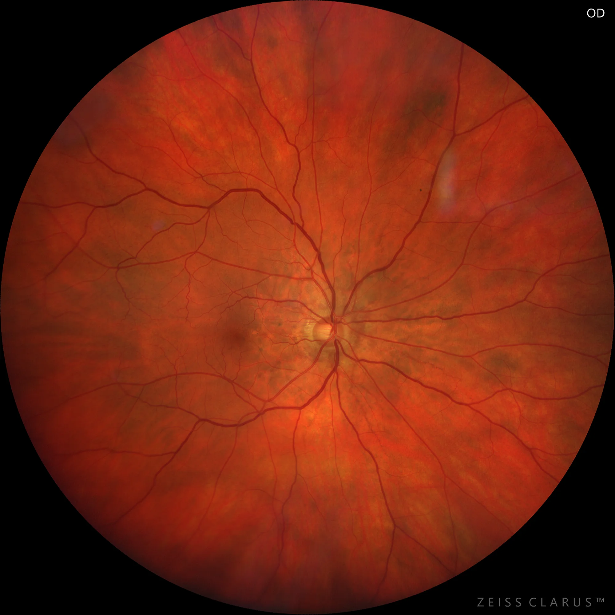
1. Right eye retinography showing slight angioid streaks.
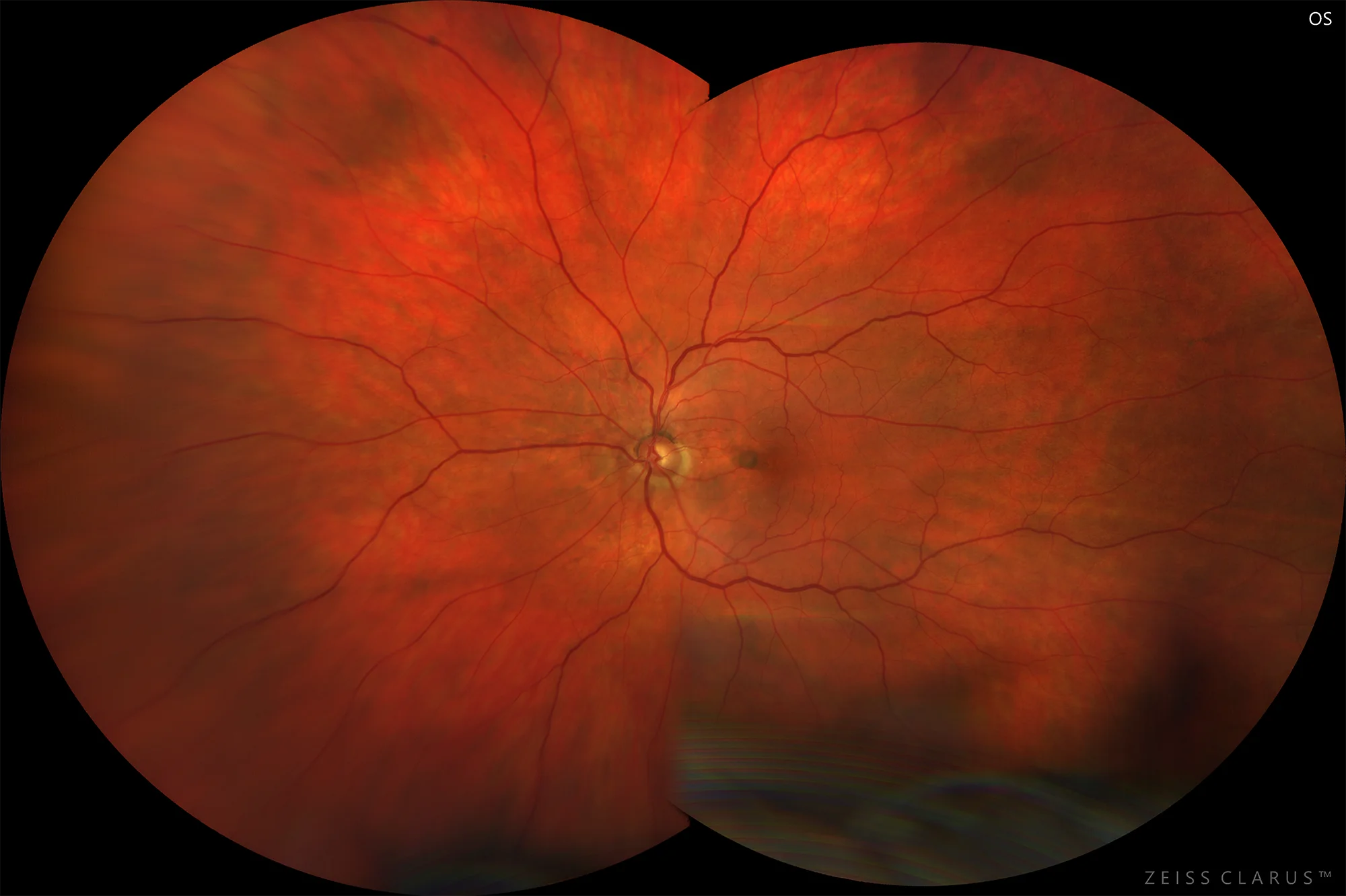
2. Left eye retinography showing angioid streaks and temporal pigmented neovascular membrane.
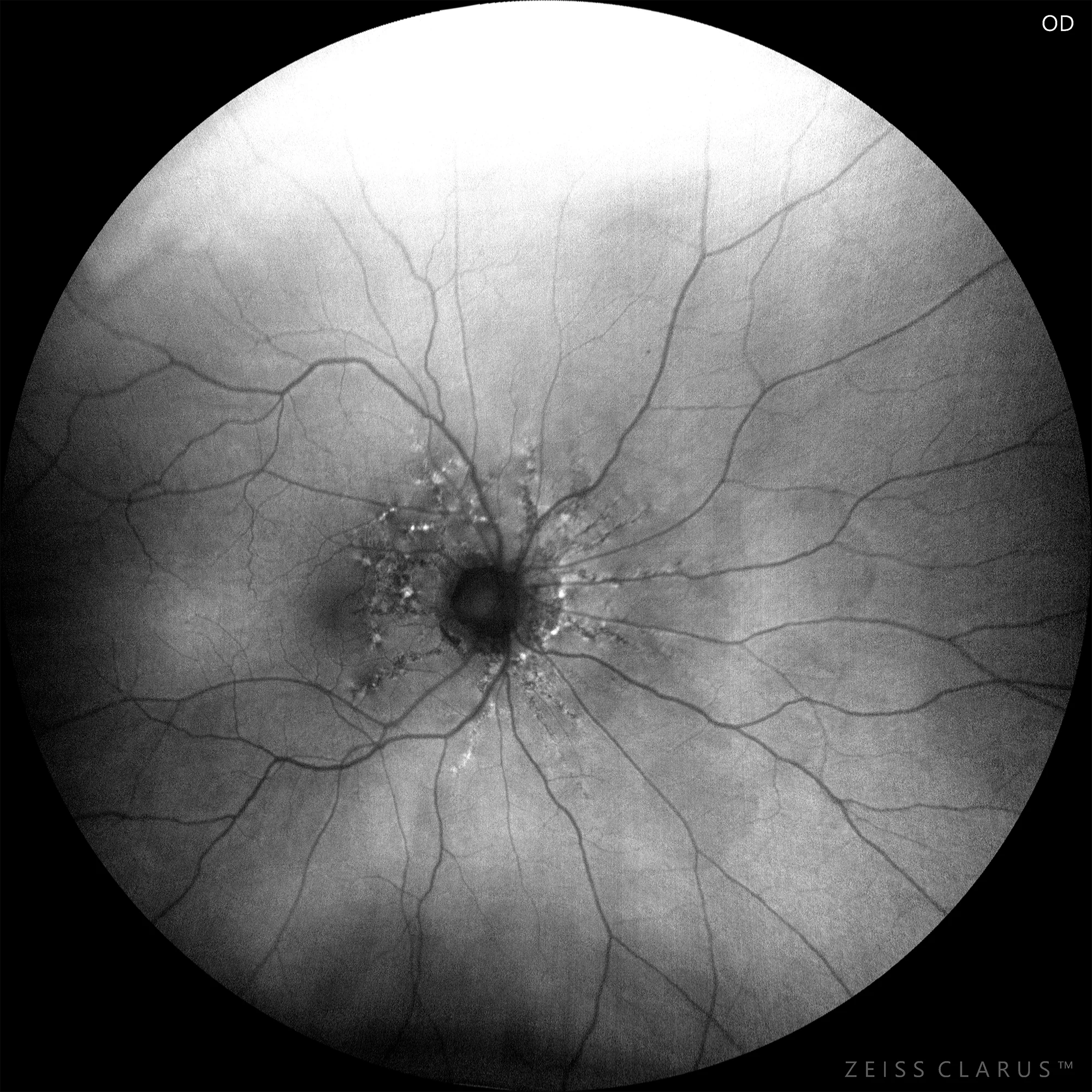
3. Right eye autofluorescence revealing angioid streaks emanating from the optic disc as areas
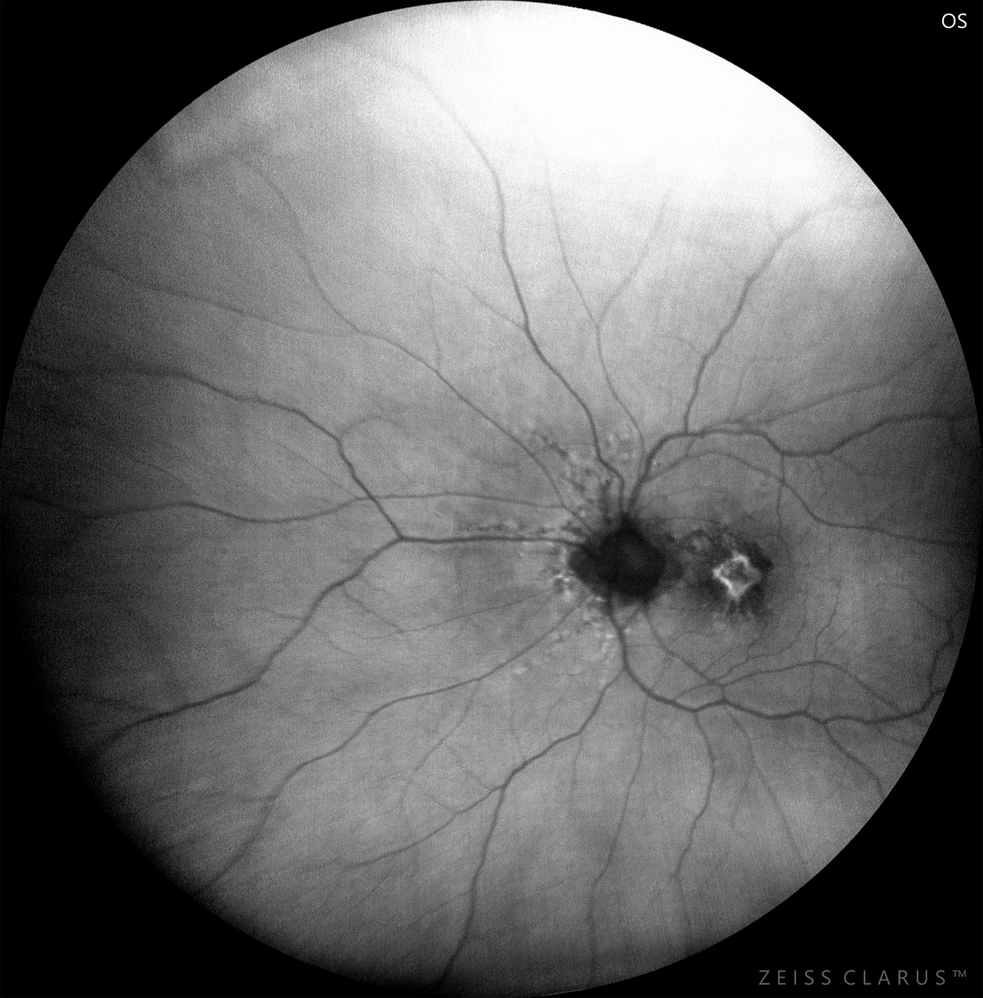
4. Left eye autofluorescence showing angioid streaks and the neovascular membrane.
Description
Angioid streaks are irregular tracts emanating from a ring of peripapillary atrophy, believed to be irregular ruptures of Bruch’s membrane. They are associated with atrophic degeneration of the retinal pigment epithelium and rupture or absence of the choriocapillaris. The most frequent complication that threatens visual acuity is the development of choroidal neovascularization in the macular region, although most patients remain asymptomatic. Angioid streaks can be associated with systemic diseases in more than 50% of cases, such as pseudoxanthoma elasticum, Paget’s disease, beta-thalassemia, sickle cell anemia, Ehlers-Danlos syndrome, among others. It is an uncommon pathology, and timely diagnosis is important, primarily in patients with family history, as well as the prevention of trauma given the potential ophthalmological complications that may arise, such as subretinal hemorrhages due to the fragility of Bruch’s membrane, as changes occur in its ultrastructure; specifically in the intermediate elastic layer and the collagenous layer.
Comments
Tests performed (equipment):
CLARUS 700:- Retinography of the right eye showing mild angioid streaks.
- Retinography of the left eye showing angioid streaks and a pigmented temporal neovascular membrane.
- Autofluorescence of the right eye revealing angioid streaks emanating from the optic disc as hypoautofluorescent areas with hyperautofluorescent borders.
- Autofluorescence of the left eye showing angioid streaks and the neovascular membrane.