Angioid streaks with CNV

Color retinography (CLARUS 500, Zeiss): Presence of angioid streaks in both eyes with marked hyperpigmentation (denoting chronicity). Small, round lesion with hyperpigmented borders in the nasal extrafoveal area corresponding to a choroidal neovascular membrane. “Orange peel” appearance temporal to the macular area.

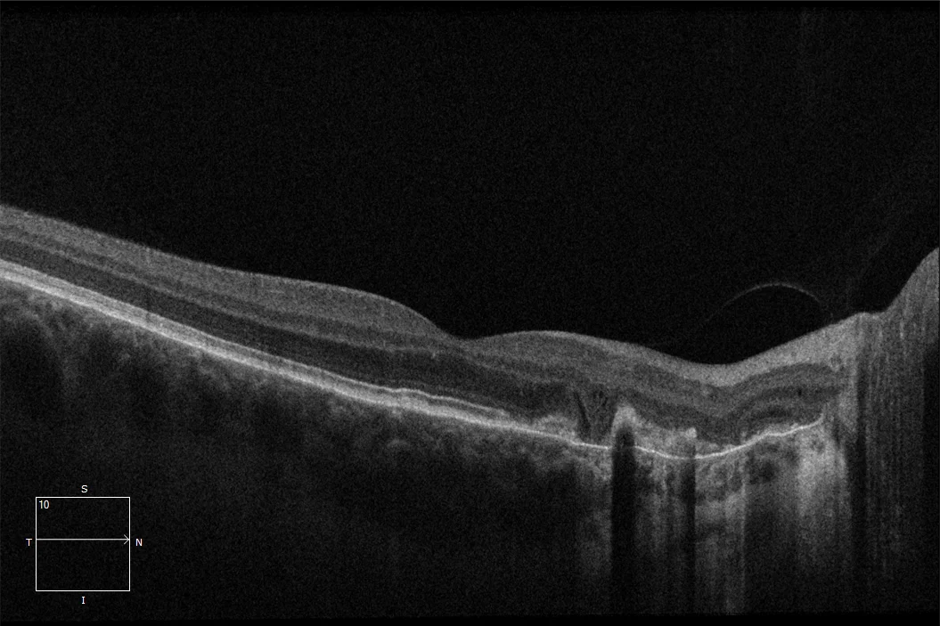
OCT (Cirrus-HD 6000, Zeiss): Extrafoveal choroidal neovascular membrane associated with angioid streaks. Disruption of outer retinal layers.
Description
Angioid streaks associated with choroidal neovascular membrane. Angioid streaks are ruptures in Bruch’s membrane that manifest in the fundus as orange or grayish bands surrounding the optic disc, extending radially from it. Various pathologies are associated with the development of angioid streaks, including pseudoxanthoma elasticum and Paget’s disease, among others. These streaks are linked to primary degeneration of elastic fibers and rupture of Bruch’s membrane. The clinical relevance of angioid streaks lies in the risk of developing late choroidal neovascularization.
Comments
Minimum attached tests: Retinography + OCT- Color retinography (CLARUS 500, Zeiss): Presence of angioid streaks in both eyes with marked hyperpigmentation (denoting chronicity). Small, round lesion with hyperpigmented borders in the nasal extrafoveal area corresponding to a choroidal neovascular membrane. “Orange peel” appearance temporal to the macular area.
- OCT (Cirrus-HD 6000, Zeiss): Extrafoveal choroidal neovascular membrane associated with angioid streaks. Disruption of outer retinal layers.