Traumatic choroidal rupture
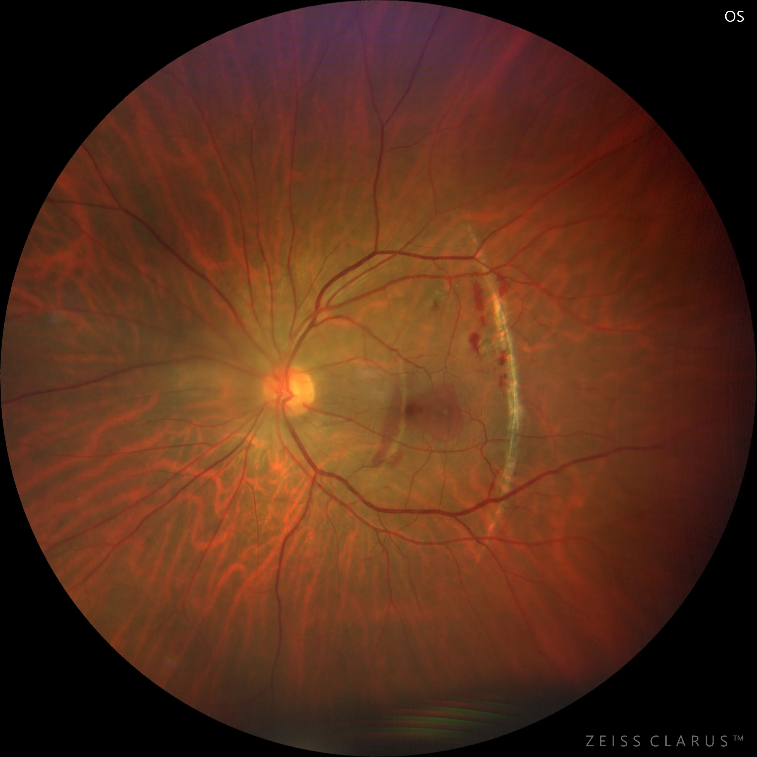
Mild hematic turbidity, with macular subretinal hemorrhages and choroidal ruptures.
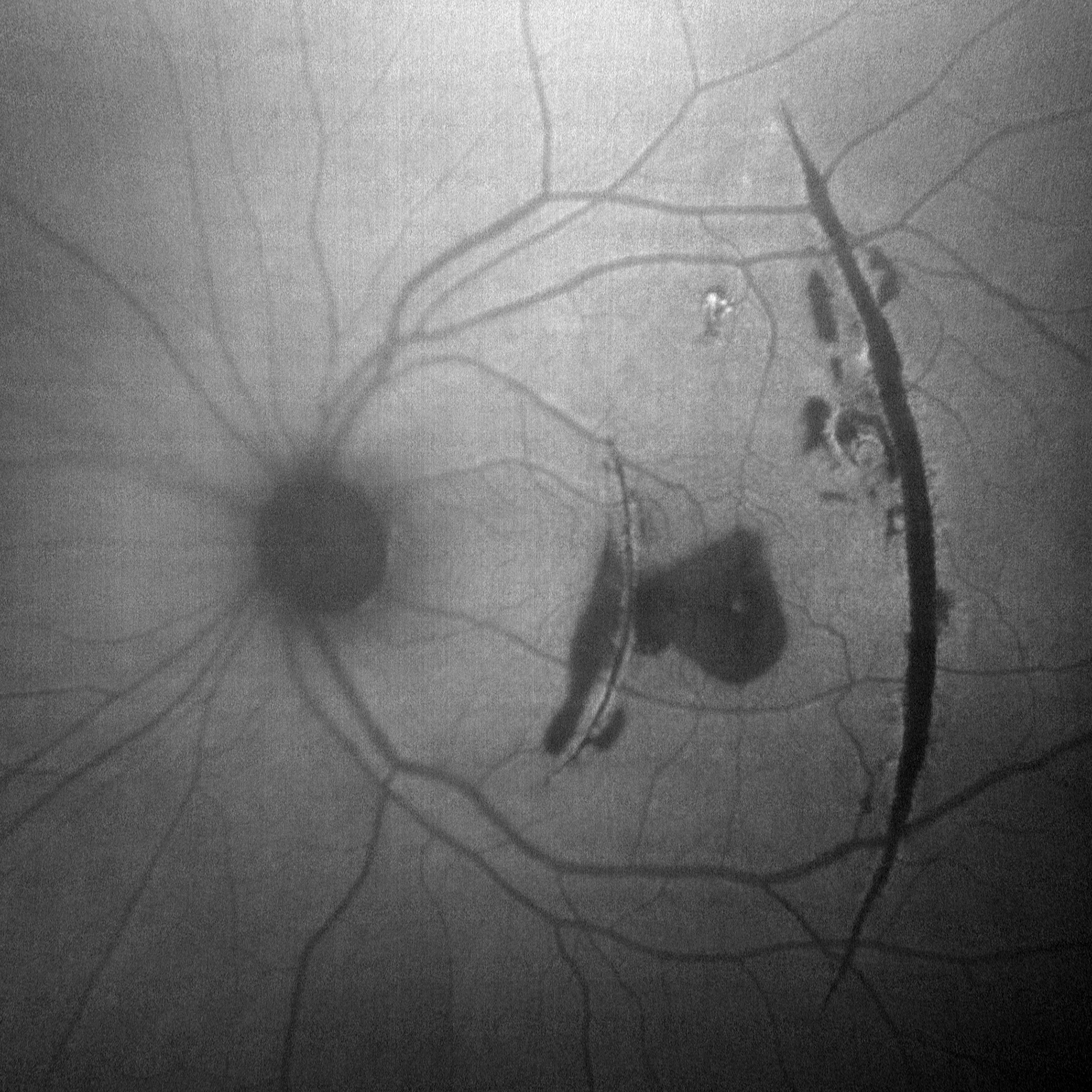
Green fundus autofluorescence (detail): Crescent-shaped hypoautofluorescent lines (choroidal ruptures), with hypoautofluorescent area due to subretinal hemorrhages
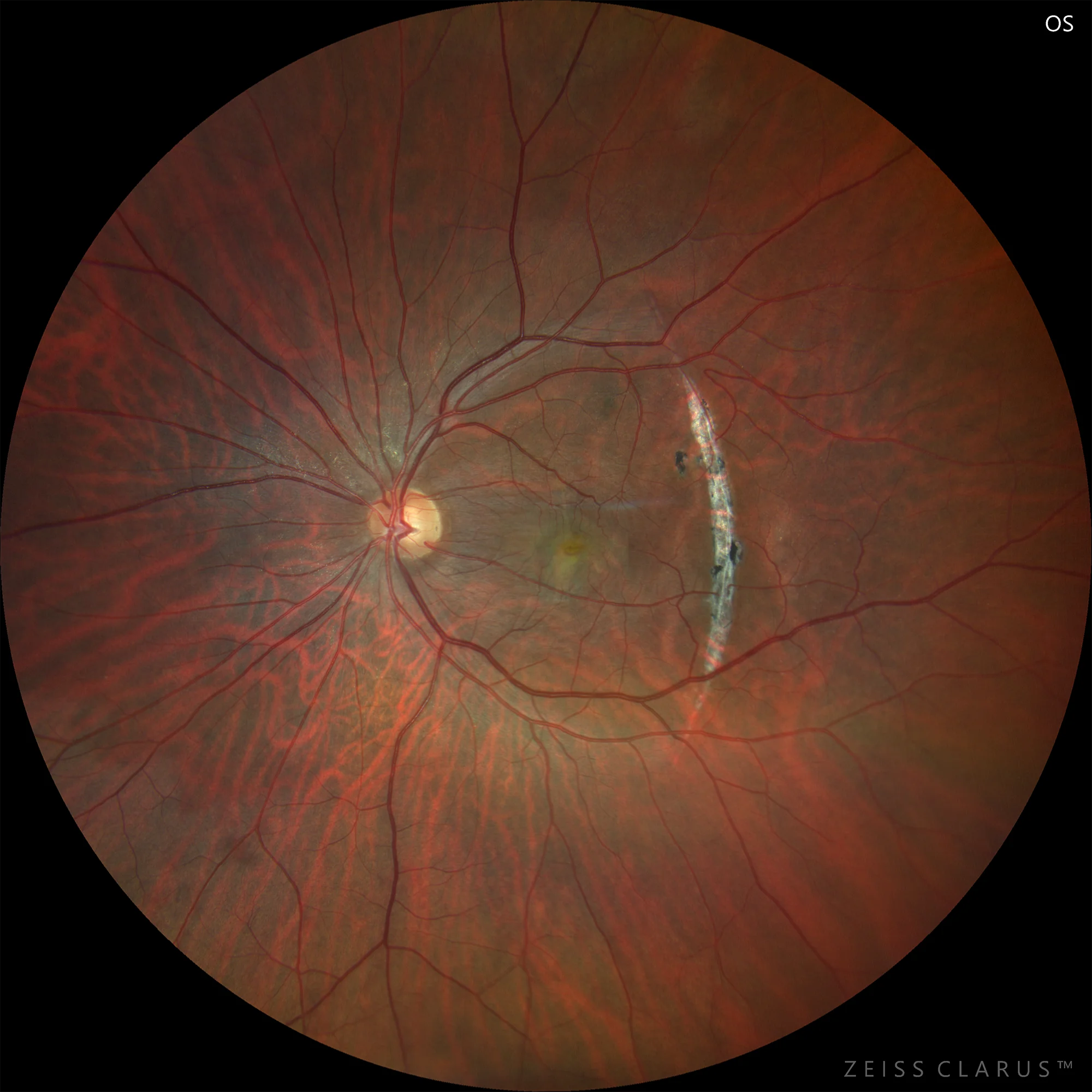
Color: Disappearance of hemorrhages with persistence of choroidal ruptures. The foveolar rupture is difficult to identify in the color image.
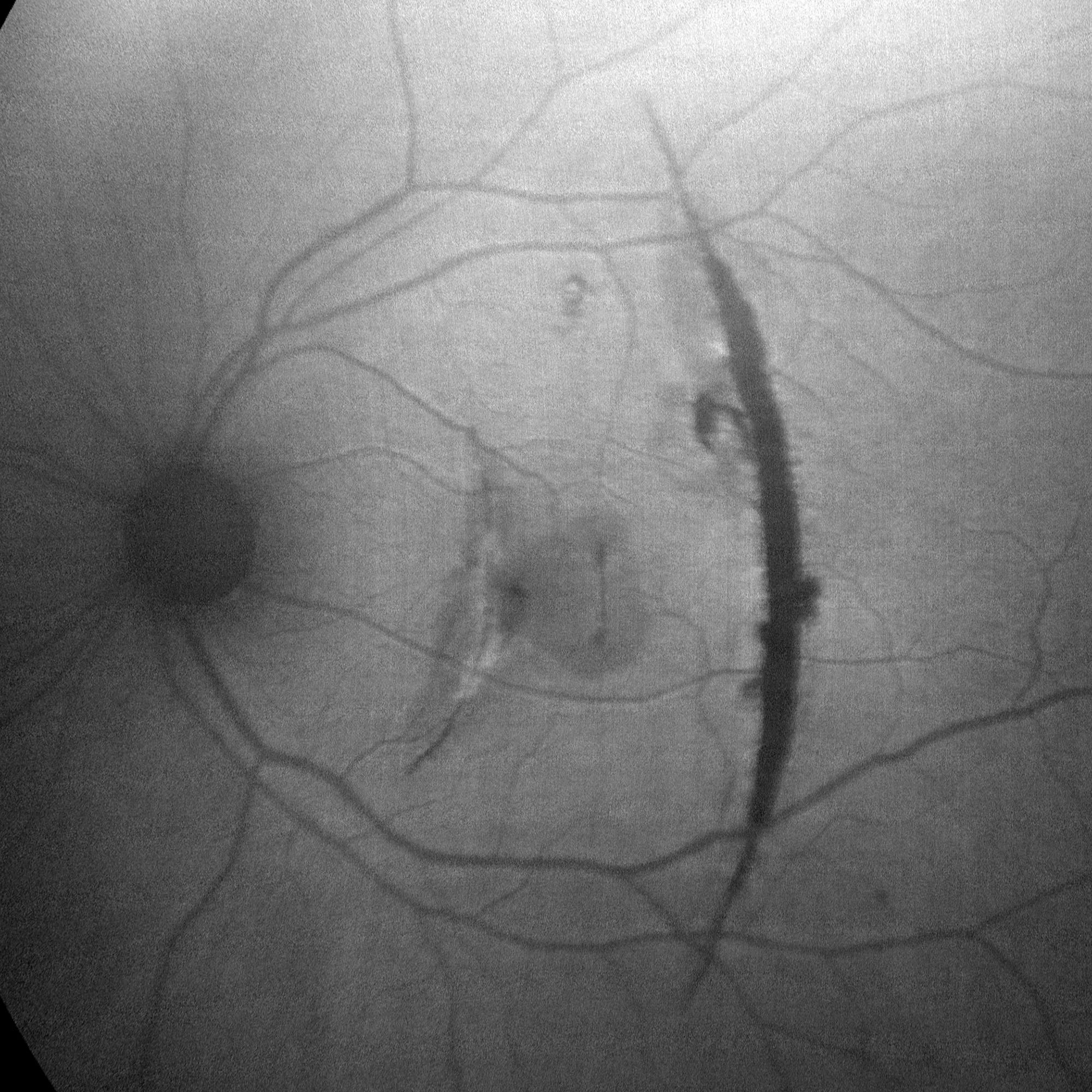
Green fundus autofluorescence (detail): Perfectly delineated choroidal ruptures as crescent-shaped hypoautofluorescent lines.
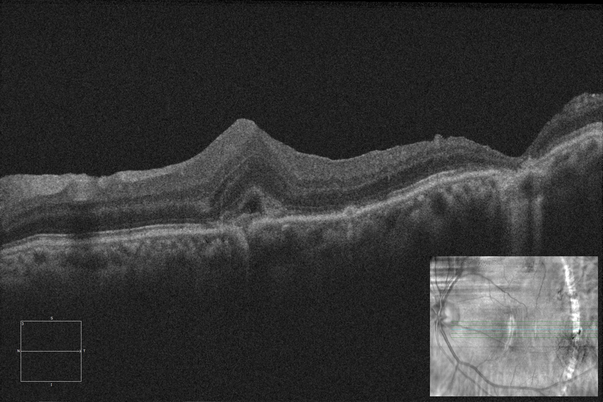
Optical coherence tomography (Cirrus-HD 5000, Zeiss): Linear OCT image with small foveolar choroidal rupture with edema and another more extensive and deep temporal rupture.
Description
Choroidal rupture occurs mainly after blunt trauma to the eyeball. It usually appears as a crescent-shaped streak concentric to the optic nerve. The visual prognosis depends on the degree of foveolar involvement, as well as the possible appearance of secondary neovascular membrane.