Sarcoid retinal vasculitis
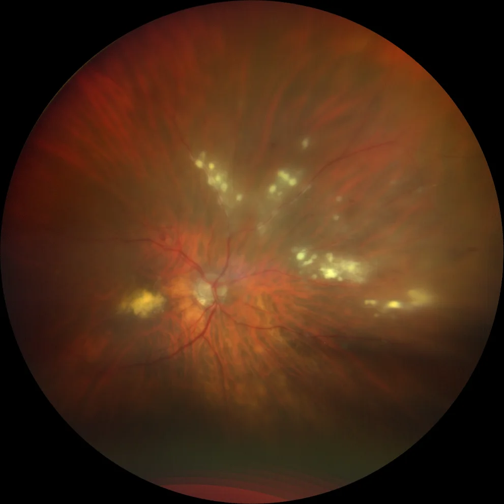
A and B. Color fundus photographs (Clarus 500, Carl Zeiss Meditec ASG, Jena, Germany) of the right and left eyes showing multiple areas of candle-wax vascular exudation, more evident in the right eye. The presence of exudates is also observed in both maculae.
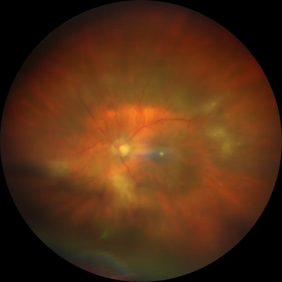
A and B. Color fundus photographs (Clarus 500, Carl Zeiss Meditec ASG, Jena, Germany) of the right and left eyes showing multiple areas of candle-wax vascular exudation, more evident in the right eye. The presence of exudates is also observed in both maculae.
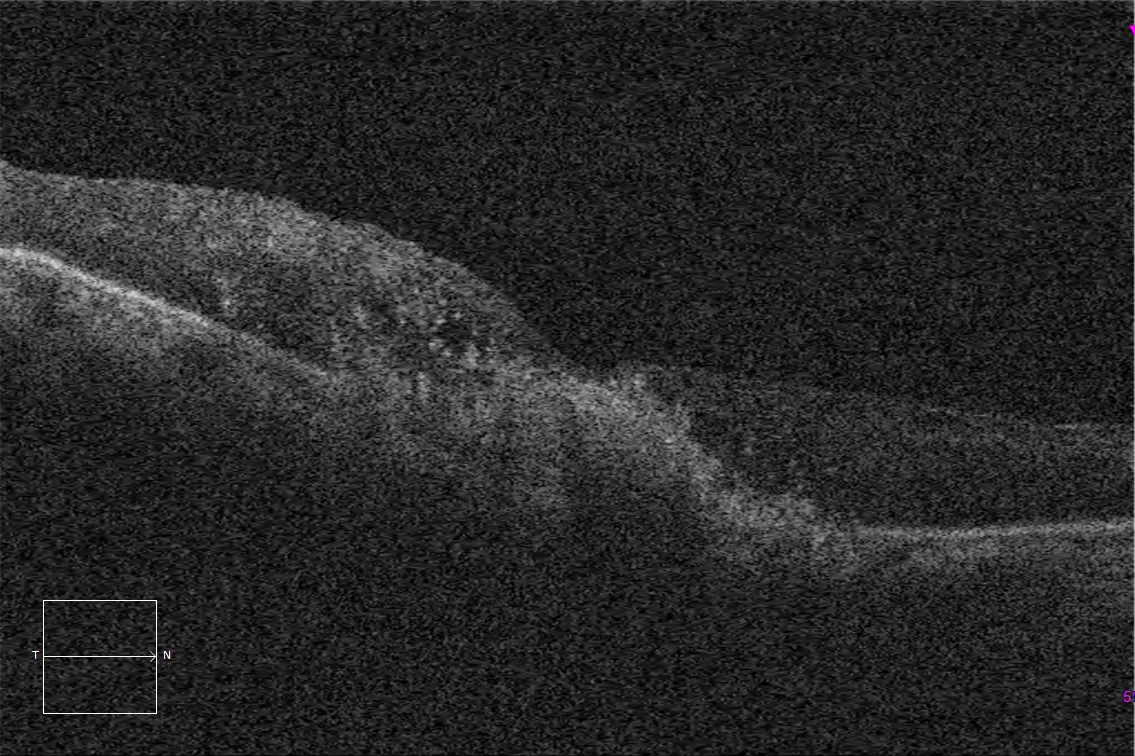
C. Macular optical coherence tomography (Cirrus 5000, Carl Zeiss Meditec ASG, Jena, Germany) of the right eye, showing the presence of subfoveal hyperrefringent exudates, as well as some intraretinal fluid cysts and hyperrefringent points at the level of the internal nuclear temporal to the fovea.
Description
Sarcoidosis is a systemic granulomatous disease with no known cause. Although thoracic involvement is its most frequent presentation, ocular involvement occurs in 10-55% of cases. Ocular inflammation in the form of uveitis is its most common ocular presentation, and can affect all ocular structures. It can present as an anterior chamber reaction classically described as retrokeratic precipitates in sheep fat, synechiae, iris nodules, vitritis, vasculitis and choroiditis. Both angiography and OCT are useful tools for its diagnosis, since macular edema is a very important cause of visual loss in these patients. Angiography shows a classic pattern of sectoral periphlebitis, with little arterial involvement that can lead to vascular sheathing. Vasculitis is usually non-occlusive, but in severe cases it can become occlusive, with the consequent risk of presenting neovascularization.