IRVAN syndrome
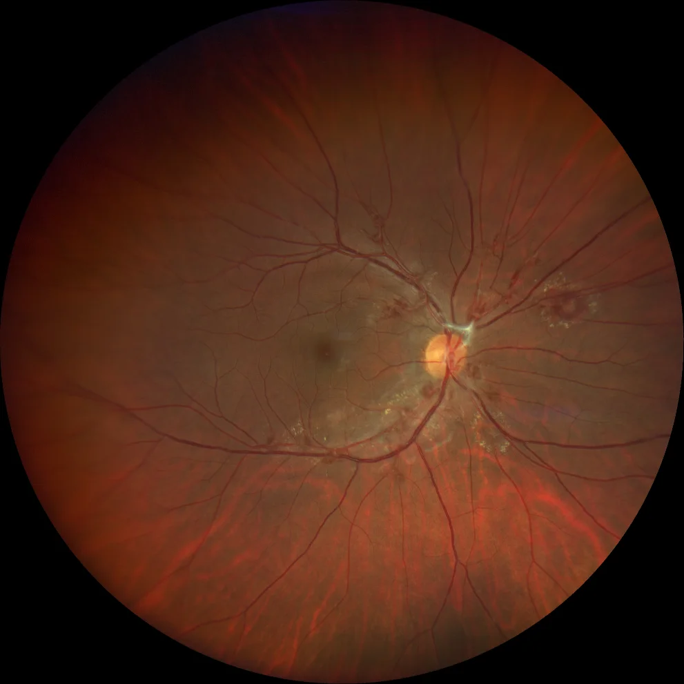
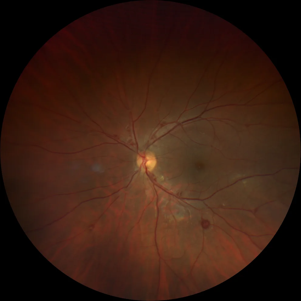
A and B. Color retinography (Clarus 500, Carl Zeiss Meditec ASG, Jena, Germany) of the right and left eyes showing aneurysms in the arterial bifurcations of the posterior pole of both eyes, some of them with lipid exudates in the surrounding circinate.
A and B. Color retinography (Clarus 500, Carl Zeiss Meditec ASG, Jena, Germany) of the right and left eyes showing aneurysms in the arterial bifurcations of the posterior pole of both eyes, some of them with lipid exudates in the surrounding circinate.
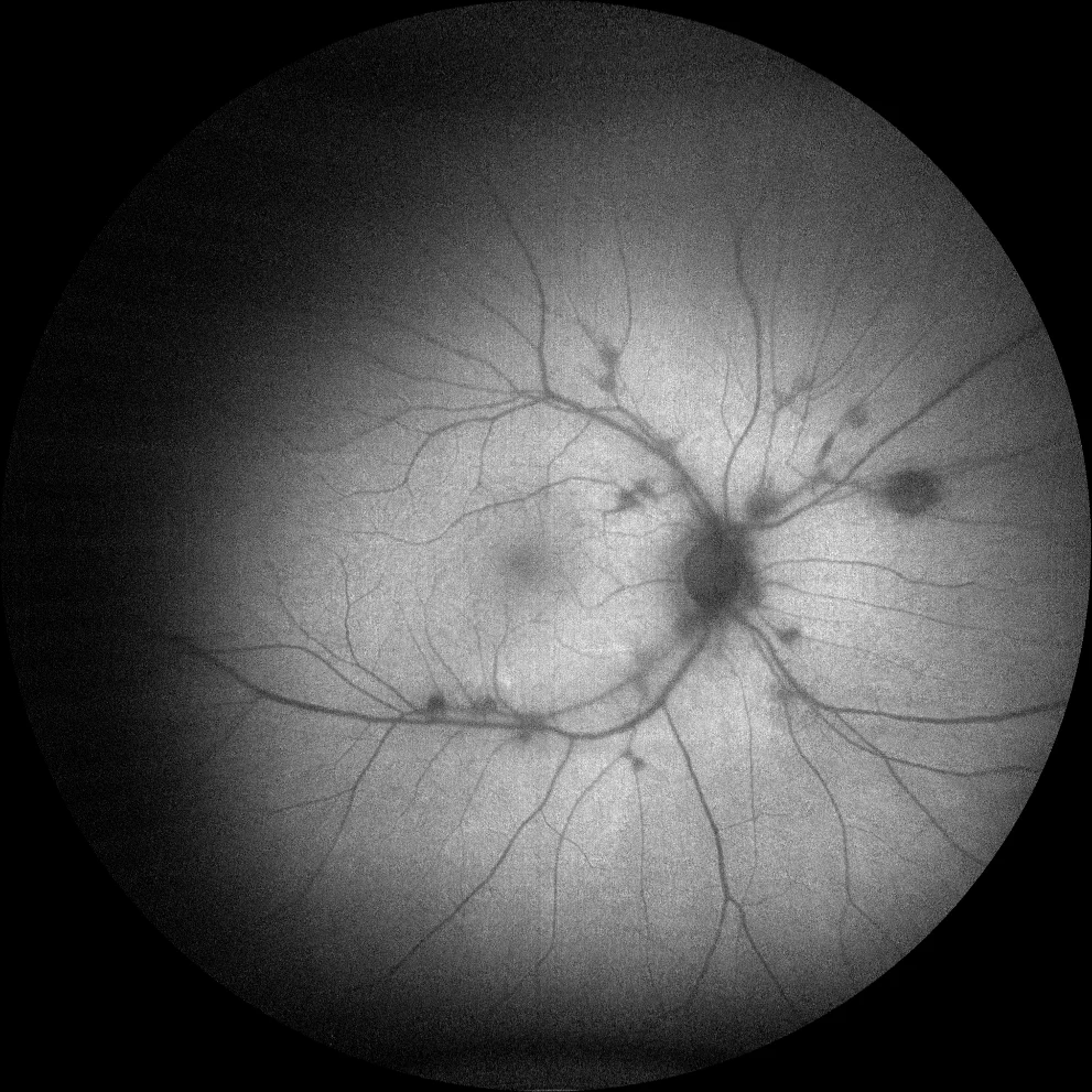
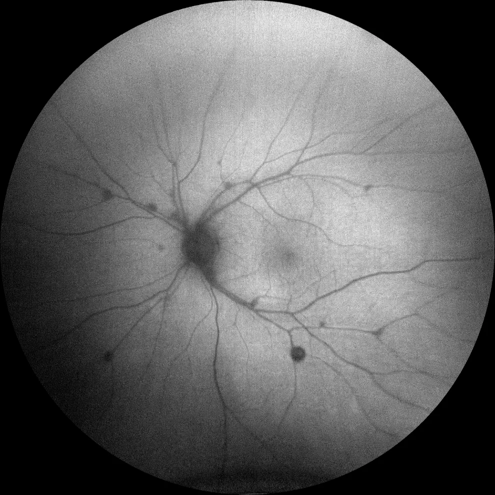
C and D. Autofluorescence (Clarus 500, Carl Zeiss Meditec ASG, Jena, Germany) of the right and left eyes, showing hypoautofluorescence in vascular lesions, due to blocking of normal retinal autofluorescence.
C and D. Autofluorescence (Clarus 500, Carl Zeiss Meditec ASG, Jena, Germany) of the right and left eyes, showing hypoautofluorescence in vascular lesions, due to blocking of normal retinal autofluorescence.
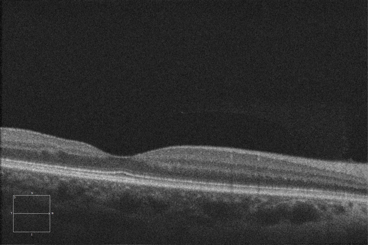
E and F. Optical coherence tomography (Cirrus 5000, Carl Zeiss Meditec ASG, Jena, Germany) of the macula, showing normal macular architecture, which explains the patient's good vision and indicates that this was a chance finding.
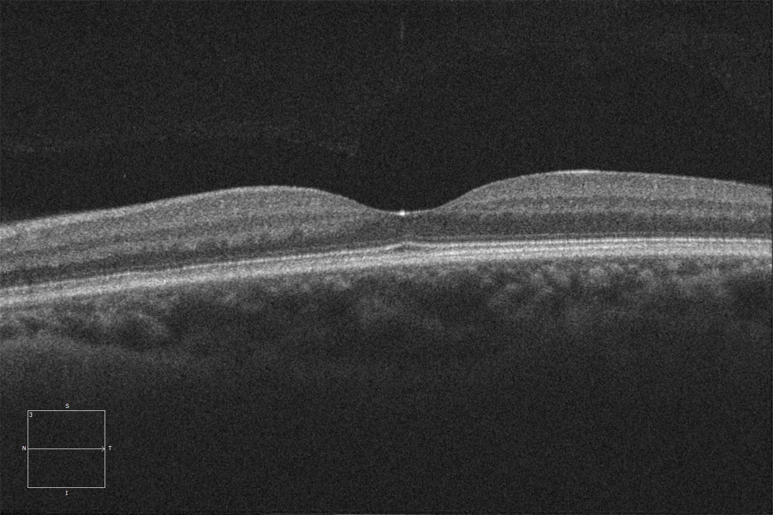
E and F. Optical coherence tomography (Cirrus 5000, Carl Zeiss Meditec ASG, Jena, Germany) of the macula, showing normal macular architecture, which explains the patient's good vision and indicates that this was a chance finding.
Description
IRVAN syndrome (Idiopathic Retinitis, Vasculitis, Aneurysms and Neuroretinitis) is a disease of unknown etiology that is not associated with any other systemic disease.
In this condition, damage to the wall of the retinal arteries is probably caused by inflammation, which promotes the appearance of macroaneurysms in the Y-shaped retinal arterial bifurcations. The diagnosis is based on a series of clinical characteristics: aneurysms in arterial bifurcations, capillary ischemia, neovascularization and exudation at the macular or peripapillary level.
Patients may present with changes in visual acuity or may be asymptomatic if the lesions do not affect the macula.
In fluorescein angiography, aneurysms are evident and fluorescein leakage can be seen from them. Capillary closure is frequently present.