< back
Idiopathic macular hole
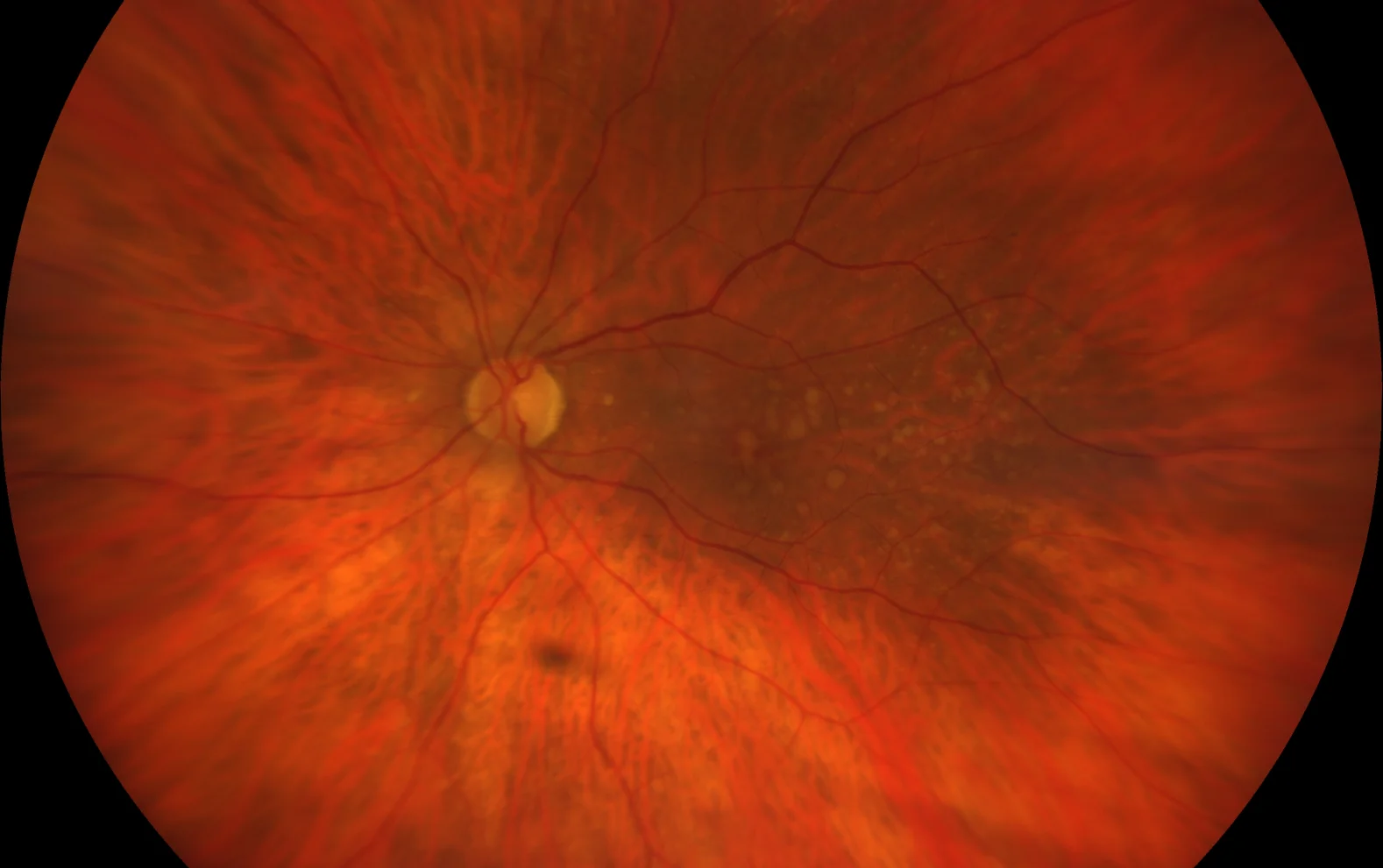
Color retinography (CLARUS, Zeiss): A whitish spot can be observed at the foveal level, corresponding to the traction zone. Presence of drusen.
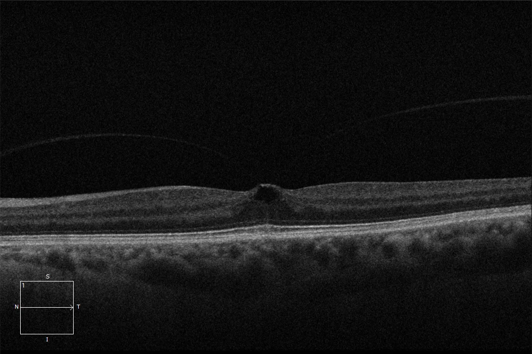
OCT (Cirrus-HD, Zeiss): Focal vitreomacular traction in the foveal area causing an alteration of the macular depression with a pseudocyst image. There is a hyperreflective line that occupies the entire foveal thickness, being the beginning of a full-thickness macular hole.
Description
An idiopathic stage I macular hole is usually asymptomatic, although it sometimes causes visual symptoms (metamorphopsia) as in the case described. In stage Ia macular holes, there is vitreofoveal traction, which causes a loss of foveal depression and pseudocysts form in the inner retina. The outer retina remains intact.