Bergmeister papilla
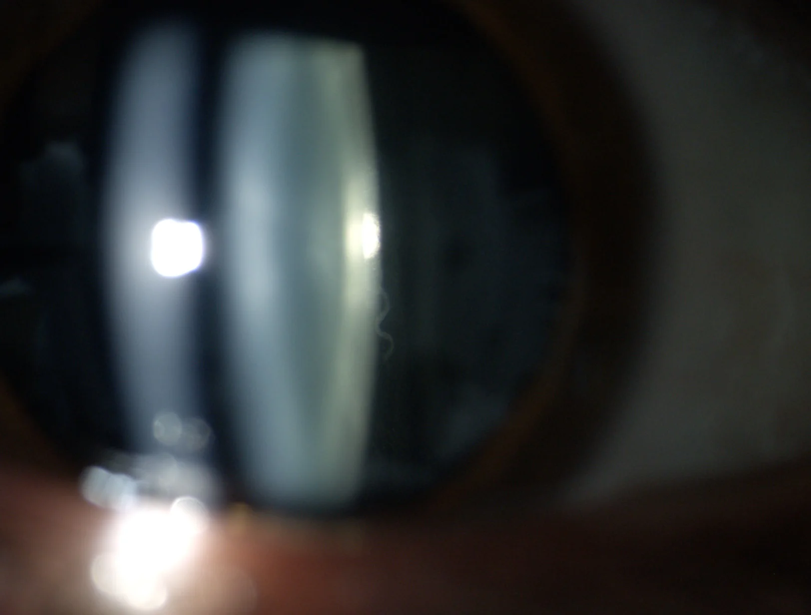
Figure 1. Remnant of hyaloid artery
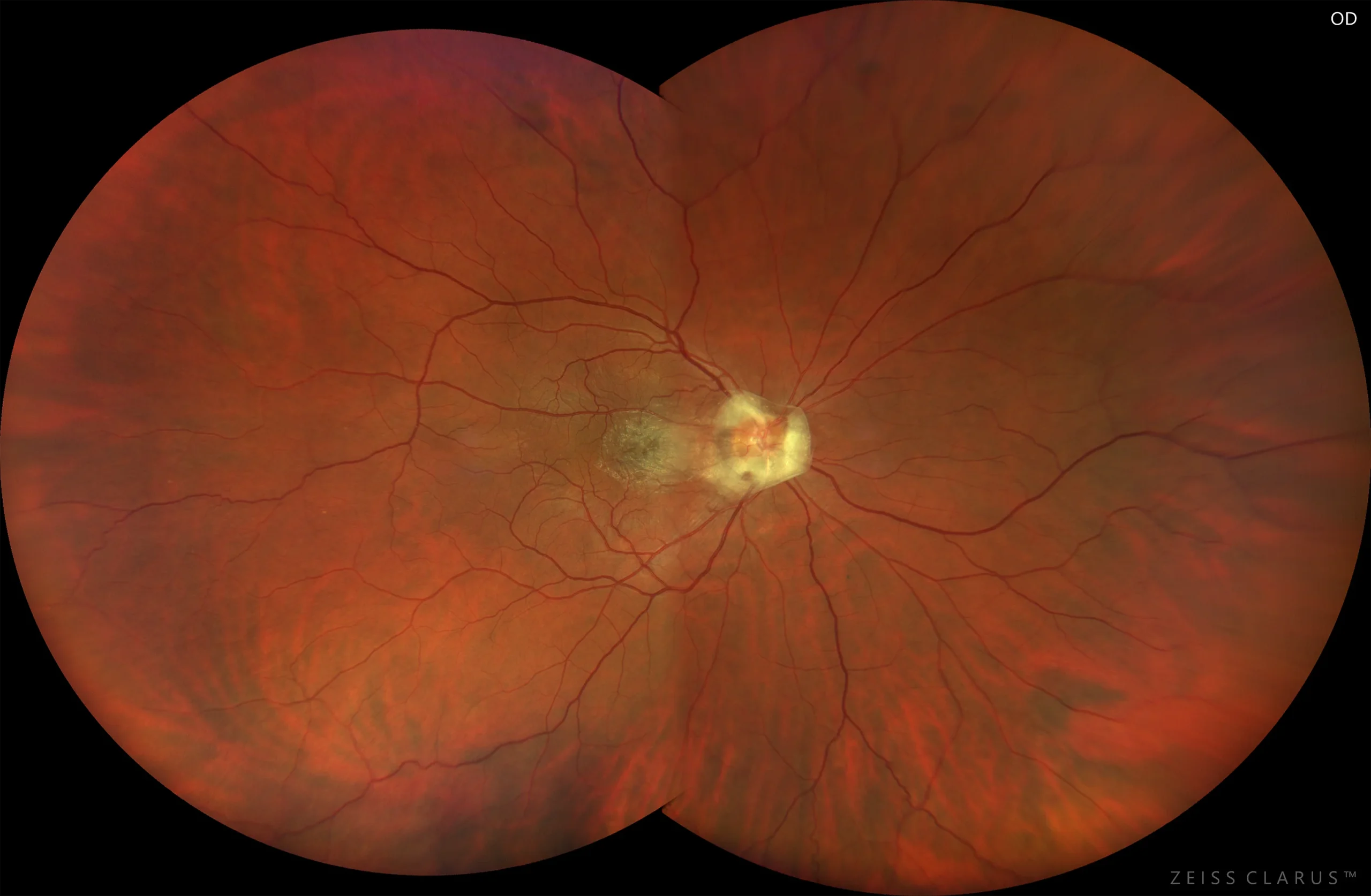
Figure 2. Color retinography of the right eye. Prepapillary glial proliferation extending to the macular area
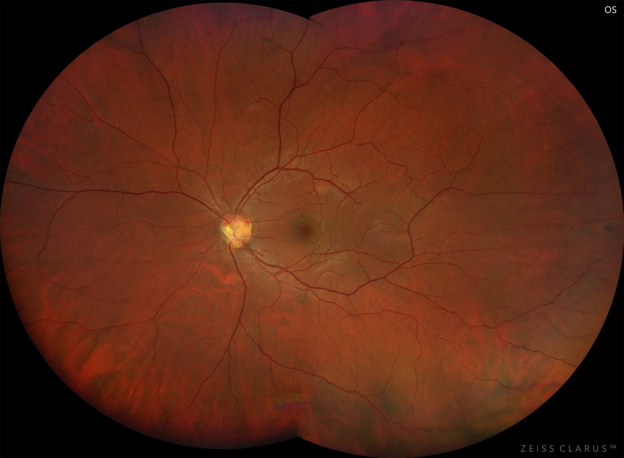
Figure 3. Color retinography of the left eye. Mild prepapillary glial proliferation.
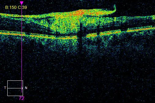
Figure 4. Prepapillary glial proliferation of the OD in OCT
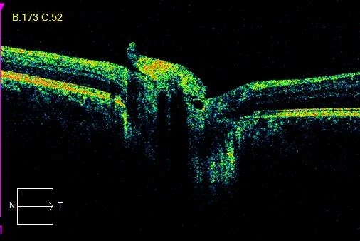
Figure 5. Prepapillary glial proliferation of the LE in OCT
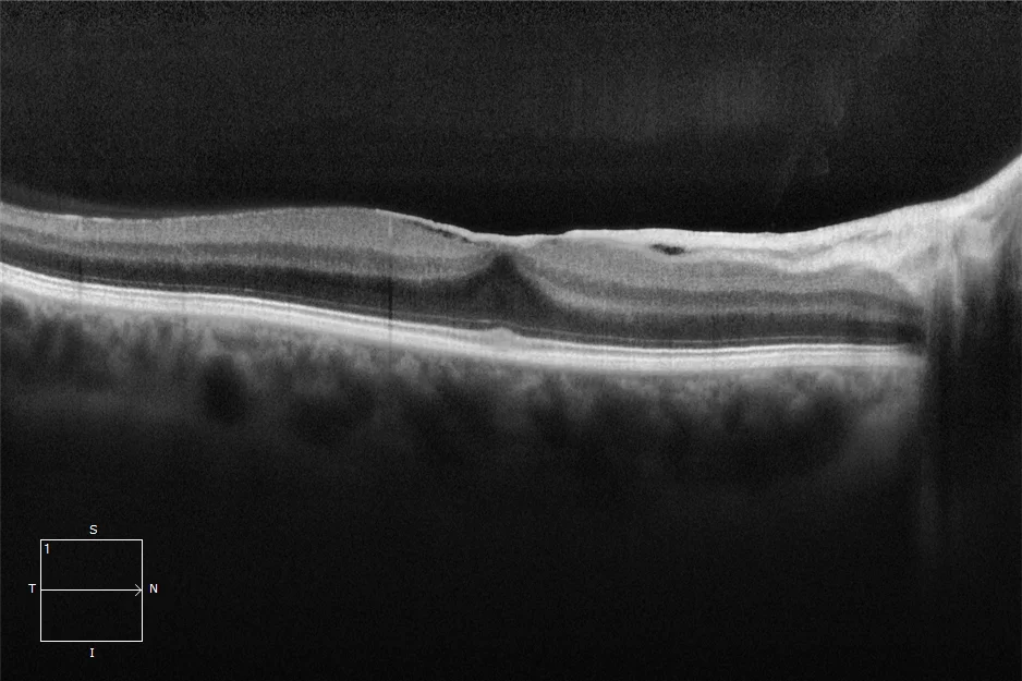
Figure 6. Extension of prepapillary glial proliferation to the macular area
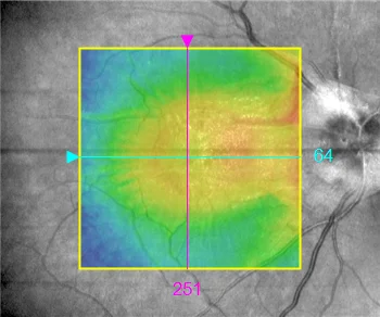
Figure 7. Macular cube. Epiretinal glial proliferation with macular thickening
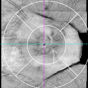
Figure 8. Prepapillary glial proliferation in en face image of the OD
Description
Bergmeister’s papilla (BP) is considered a remnant of the persistent hyaloid artery at the level of the optic disc, and appears as an epipapillary membrane that can occlude the papilla or part of it. It is usually unilateral and without clinical repercussions, often being a chance finding. The embryonic hyaloid artery, a branch of the primitive dorsal ophthalmic artery, is responsible for irrigating the lens during embryonic development and is progressively reduced from the tenth week of gestation until it disappears at birth. The incomplete disappearance of these embryonic structures on the head of the optic nerve causes the permanence of glial tissue at the beginning of the Cloquet canal or Stilling duct that connects the optic nerve with the lens, crossing the vitreous humor and which would be the complete remnant of the hyaloid artery.