Persistence of myelin fibers
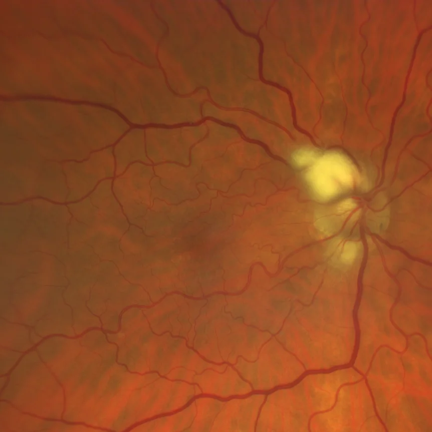
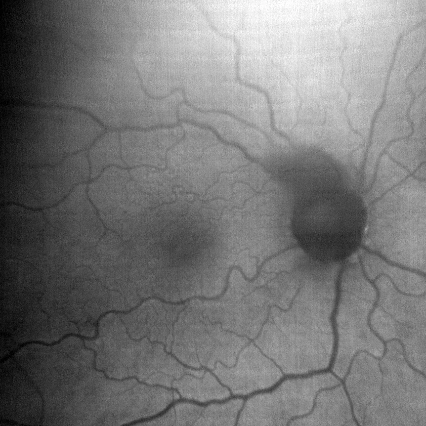
Color retinography (detail, Clarus 700, Zeiss). Whitish peripapillary lesion at 9 to 12 hours and fainter at 6 hours in RE.
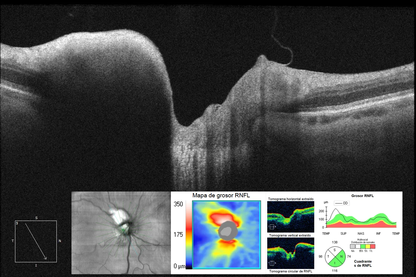
Optical coherence tomography (Radial and Optis disc cube, Cirrus-HD 5000, Zeiss): Increased thickness and reflectivity of the RNFL located 10 to 12 hours peripapillary, with shadow effect on the outer retinal layers.
Description
Persistence of myelinated nerve fibers is a congenital abnormality that is usually asymptomatic, unilateral or bilateral, and appears as whitish streaks, usually close to the optic disc. Diagnosis by fundus examination is usually simple. Optical coherence tomography shows thickening of the nerve fiber layer (RNFL) with increased signal and shadowing of the outer retinal layers, which should not be confused with true optic disc edema.