< back
Retinal astrocytic hamartoma
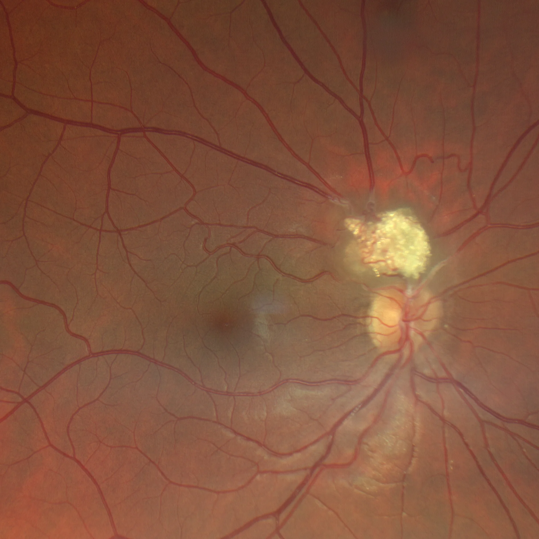
Color retinography and AF: Calcified and vascularized multinodular lesion, with positive autofluorescence.
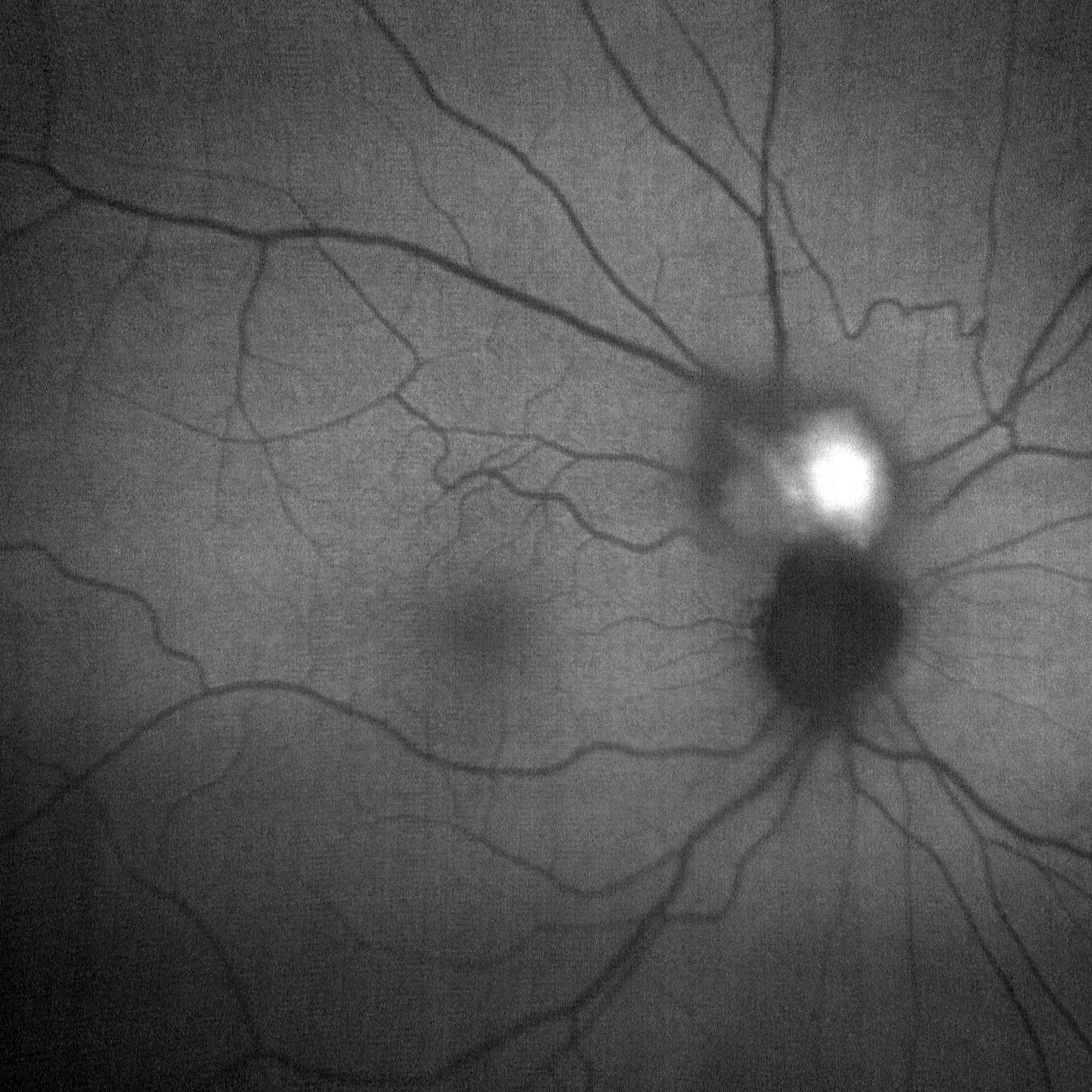
Color retinography and AF: Calcified and vascularized multinodular lesion, with positive autofluorescence.
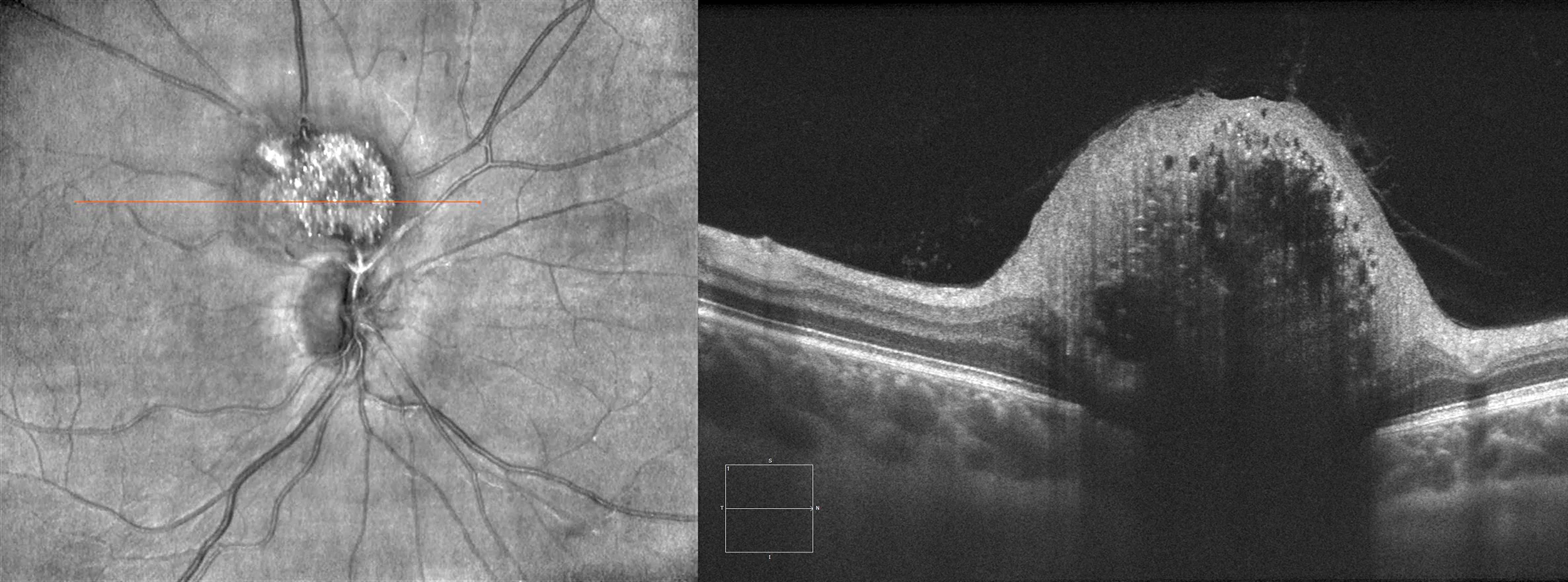
OCT 1 line HD: Hyperreflective dome-shaped lesion with a moth-eaten appearance with a marked posterior shadow.
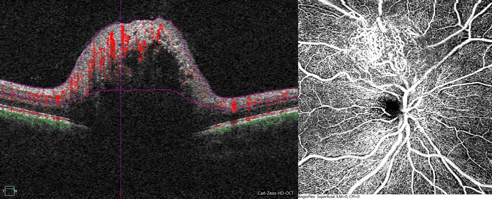
OCTA: Tortuous tumor vascularization
Description
Retinal astrocytic hamartoma
Retinal astrocytic hamartoma is a glial, vascularized, and benign tumor, usually asymptomatic. It typically presents as a flat or multinodular whitish lesion, single or multiple, frequently associated with tuberous sclerosis.