< back
Advanced chronic simple glaucoma
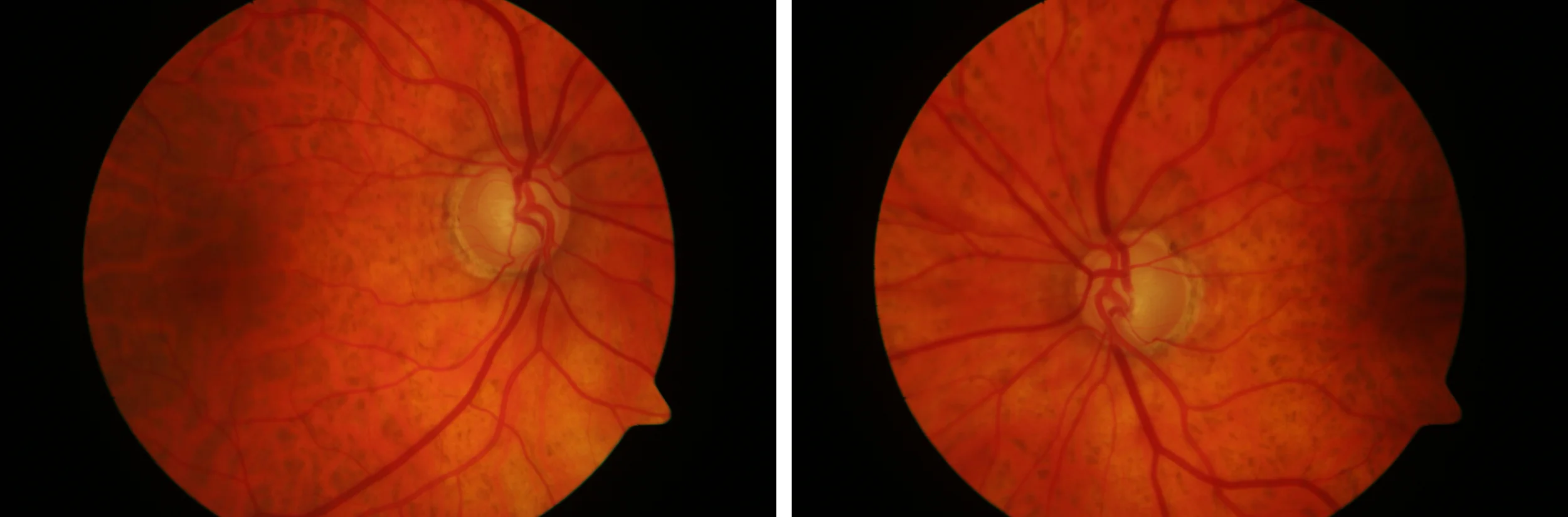
Color retinography 30º: Tiger-colored fundus. Papillae with 0.9 excavation and marked loss of neuroretinal ring.
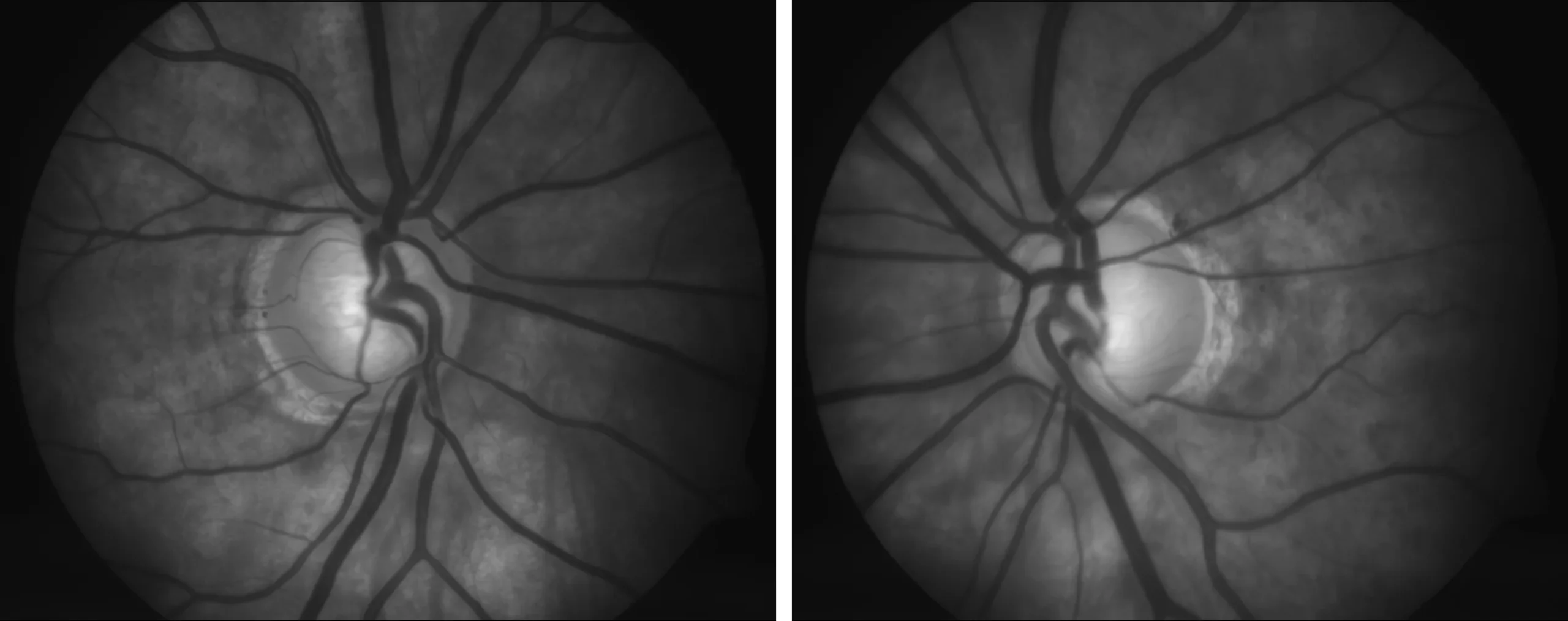
Retinography with 20º green filter: Allows better visualization of the details of the papilla, highlighting the bayonet-shaped retinal vessels and the nasal displacement of large vessels.
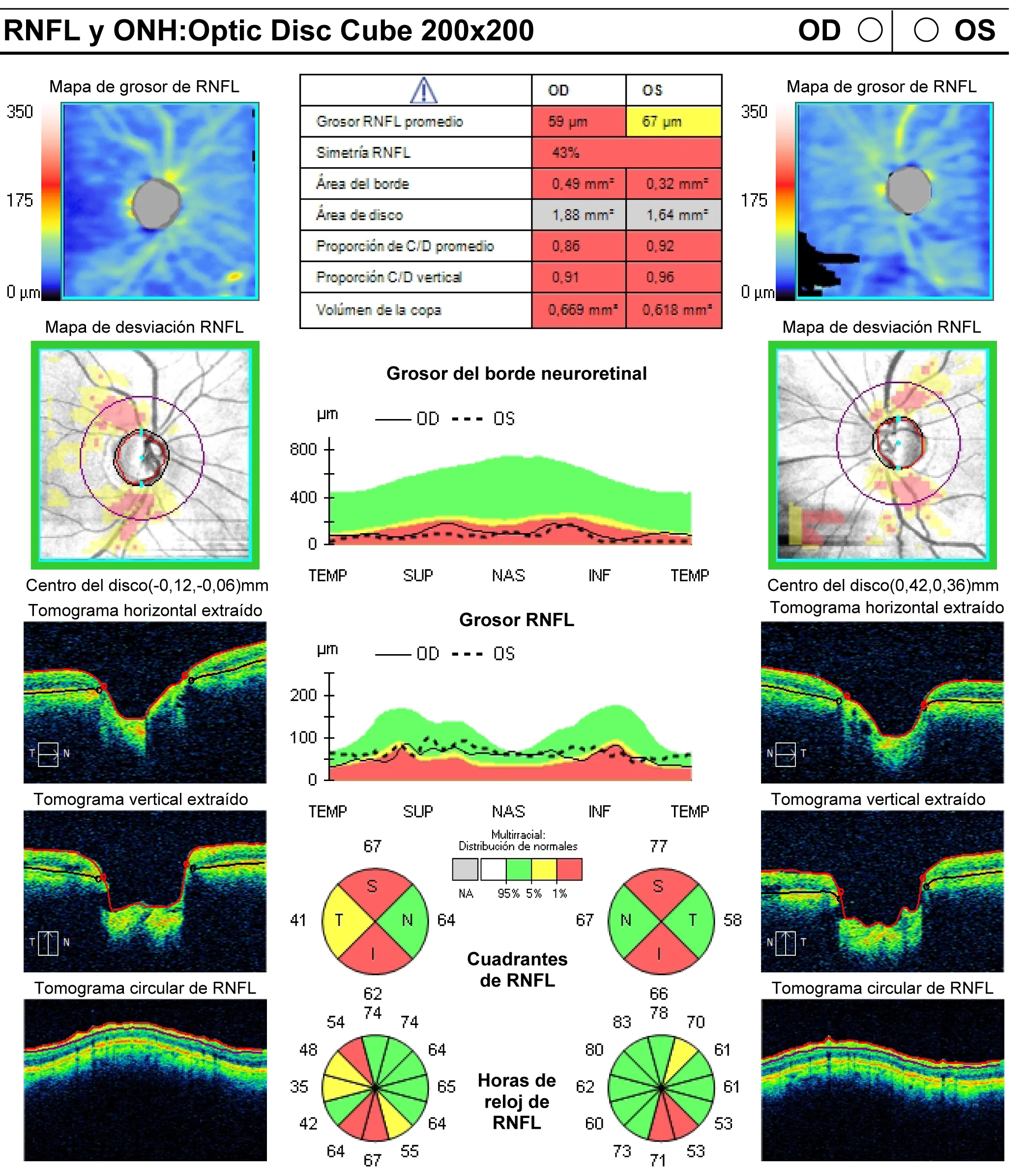
Papillary OCT (RNFL)
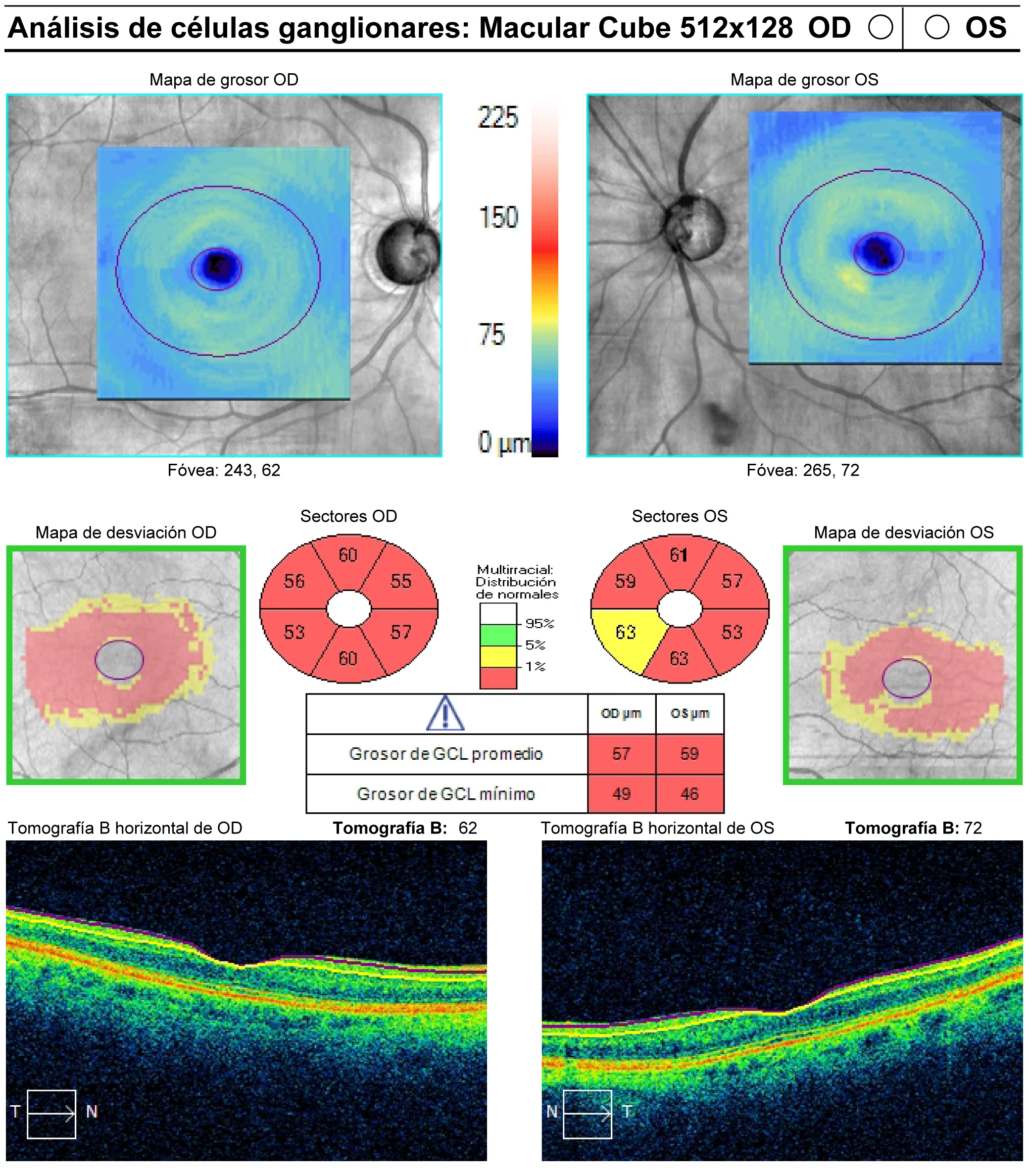
Macular OCT: Ganglion cell layer analysis
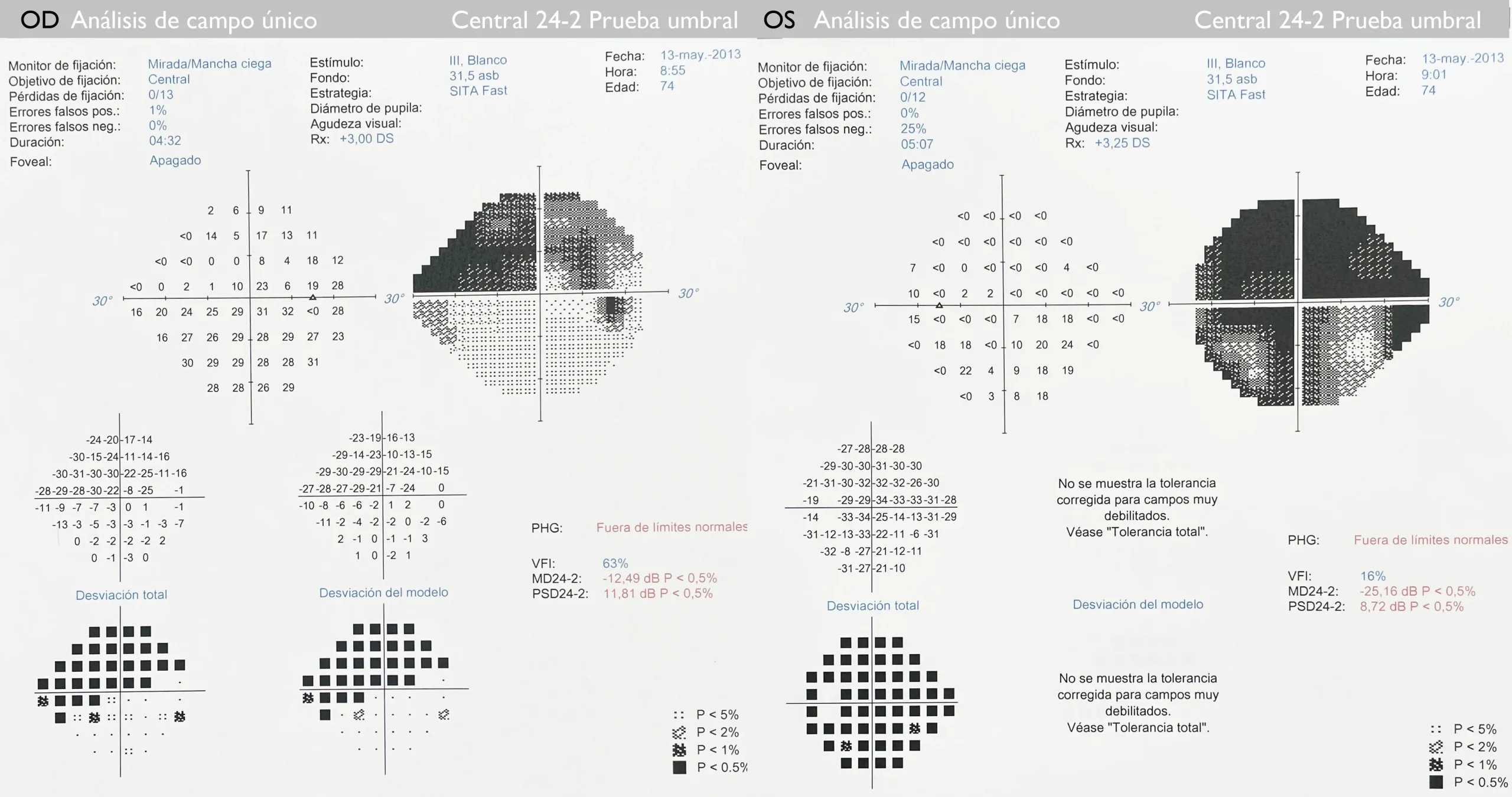
Visual field (threshold test 24.2 Humphrey, Zeiss): Marked CV involvement: OD: Upper arcuate defect with nasal step. LE: subtotal loss.
Description
Chronic simple glaucoma (CSG) is a progressive optic neuropathy secondary to increased intraocular pressure that causes visual field loss that can lead to blindness.