Occult optic nerve drusen
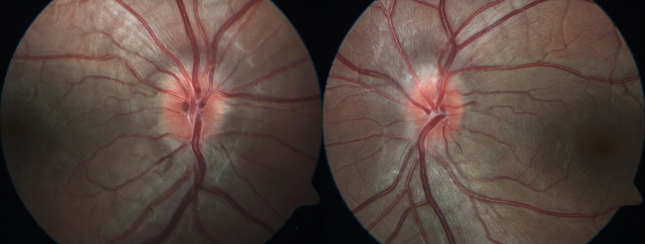
Color retinography (30º, FF 450 IR plus, Zeiss): Raised hyperemic papillae with poorly defined edges in the nasal and upper AO areas (pseudopapilledema). Increased vascular branching.
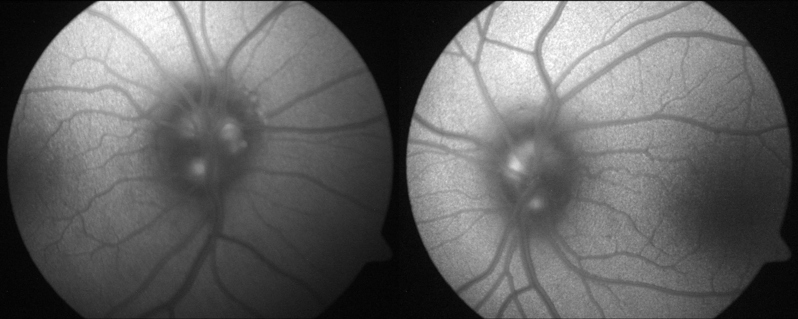
Background autofluorescence (30º, FF 450 IR plus, Zeiss): Positive autofluorescence (confirms presence of hidden drusen).
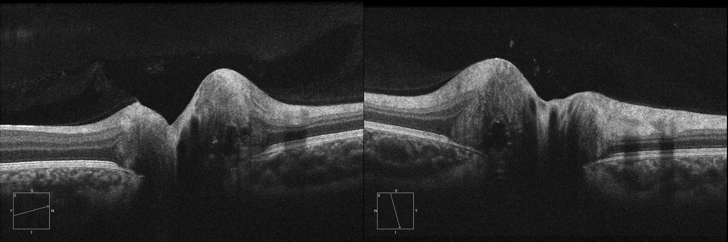
Optical coherence tomography (EDI radial OCT, Cirrus-HD 5000, Zeiss): Raised papilla with small rounded hyporeflective formations with hyperreflective edges, characteristic of optic nerve drusen.
Description
Optic nerve drusen are deposits that calcify over the years, usually asymptomatic and bilateral. In children, they produce an elevated optic nerve with poorly defined edges that can be confused with true papilledema. Diagnosis is usually confirmed by ultrasound, optical coherence tomography (OCT) and fundus autofluorescence.