Tractional diabetic retinopathy
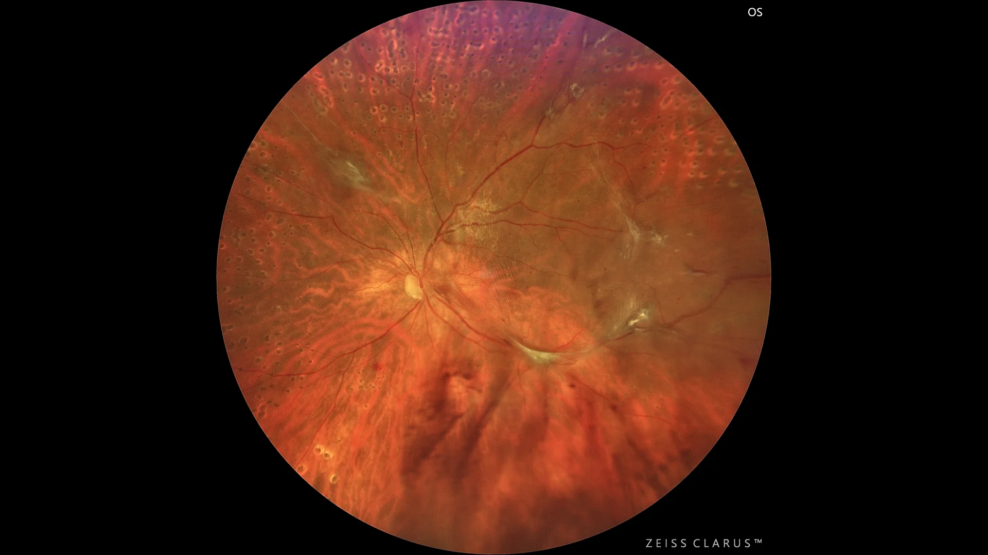
Color retinography, in which diabetic fibrovascular proliferation is observed, establishing a connection between the papilla and the temporal vascular arcades, with evident signs of traction.
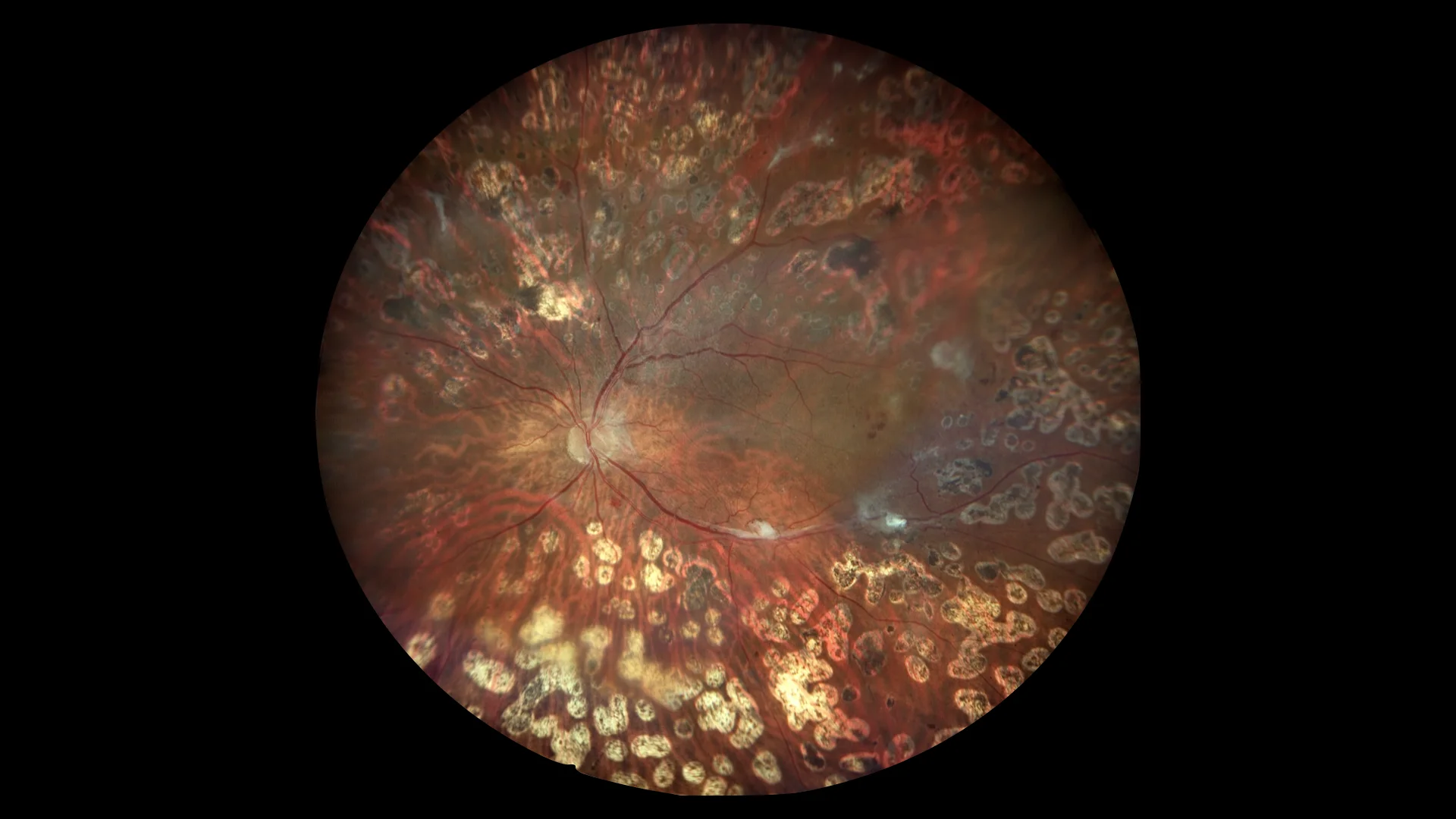
Post-surgery color retinography: In this image we see fibrosis of the neovascular tufts, as well as their segmentation.

Color retinography in a patient with hemovitreous and tractional retinal detachment, in which vitreoretinal proliferation can be seen pulling on the posterior pole.
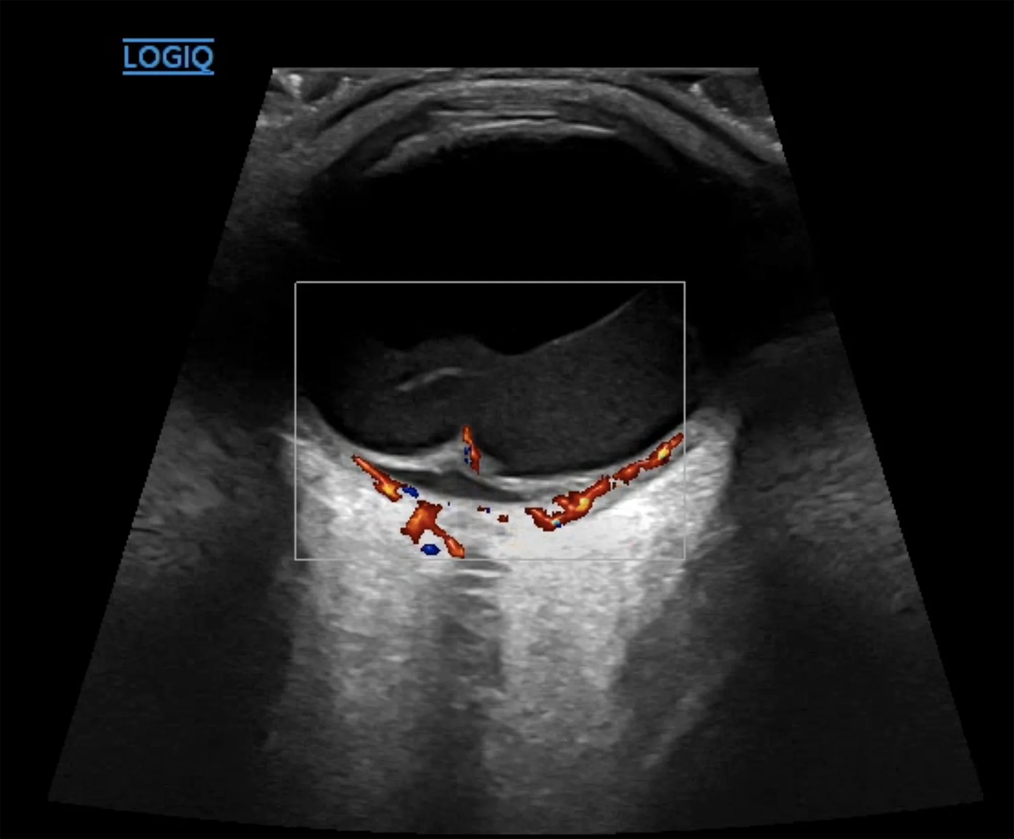
Doppler ultrasound showing the traction of the proliferative membranes and the flow within them. The hyperreflective content in the vitreous cavity secondary to the hemovitreous can also be seen.
Description
Tractional diabetic retinopathy . Tractional diabetic retinopathy is a severe form of diabetic retinopathy that occurs when abnormal blood vessel growth in the retina leads to the formation of fibrous tissue. This tissue can shrink and cause traction on the retina, resulting in detachment and possibly severe vision loss. The main treatment is vitrectomy, a surgery to remove the fibrous tissue and relieve the traction. Prevention through strict blood sugar control and regular eye exams is key to avoiding this complication.