Tractional retinal detachment
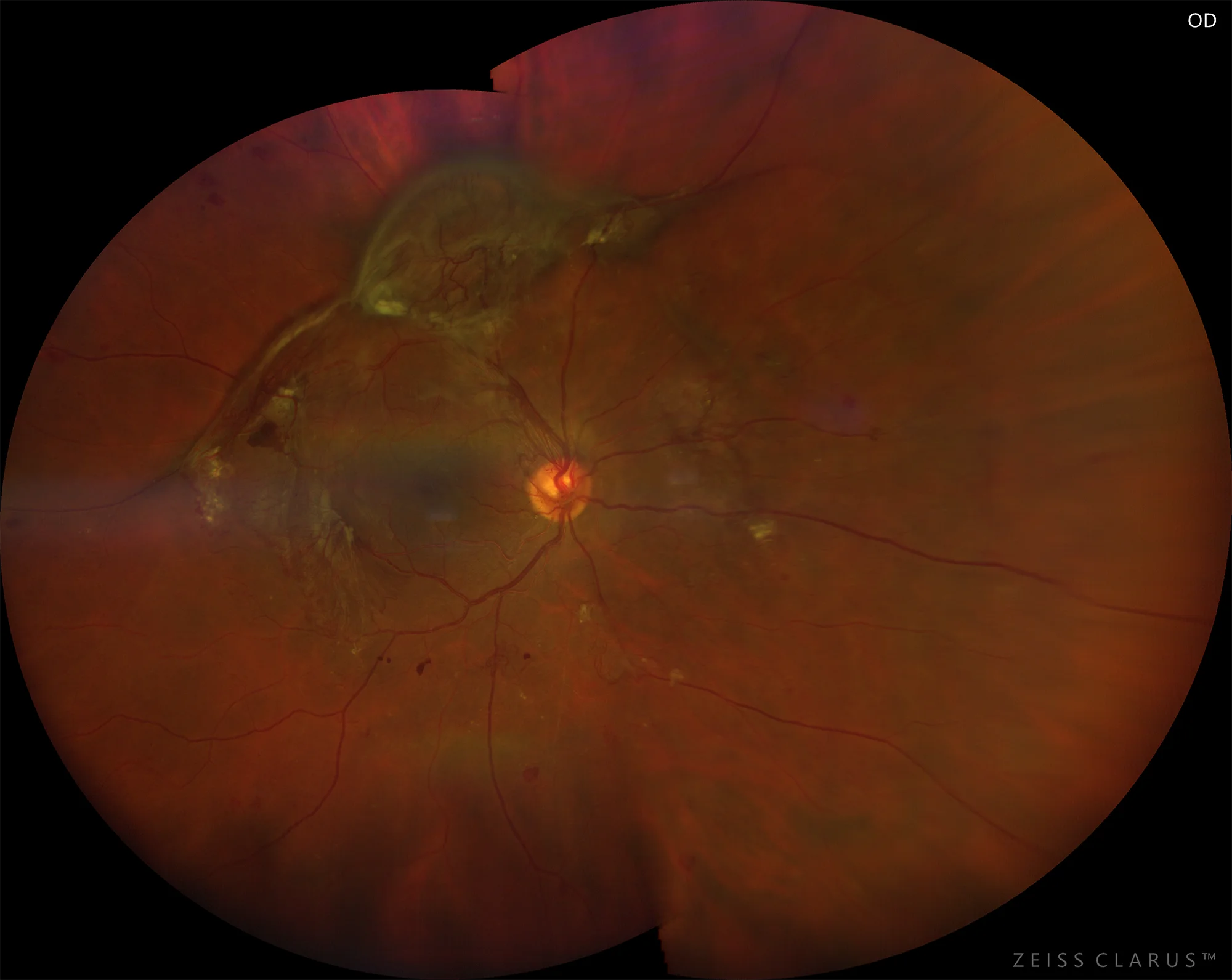
Figure 1. Color retinography showing the upper tractional retinal detachment of the right eye
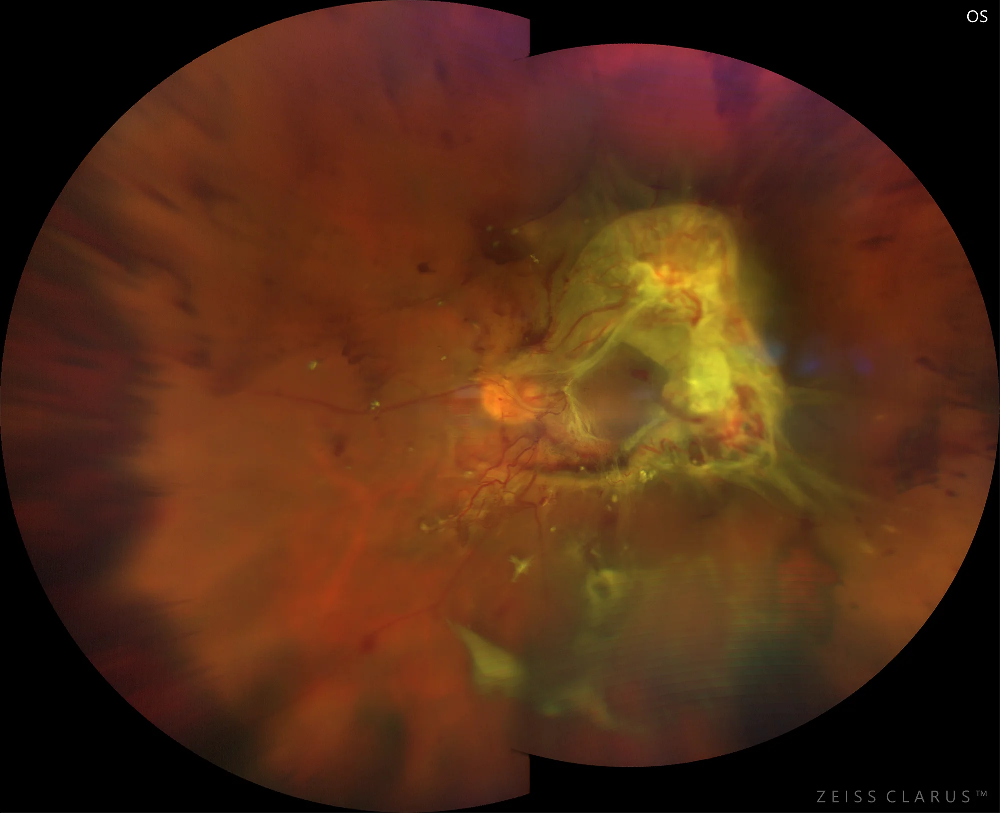
Figure 2. Color retinography showing central tractional retinal detachment of the left eye
Description
Tractional retinal detachment (RD) can occur secondary to penetrating injuries or proliferative vascular retinopathies. These generate traction on the neurosensory retina that causes it to separate from the retinal pigment epithelium (RPE). In some cases, the traction can cause a retinal tear and thus produce a rhegmatogenous and tractional retinal detachment. Clinically, the retina loses its concavity and assumes a convex shape, producing a progressive lifting of the retina, which is accompanied by progressive and irreversible visual loss in advanced stages. Treatment consists of releasing the traction by performing a vitrectomy, although the functional prognosis is usually very limited.