Macular telangiectasias type 2
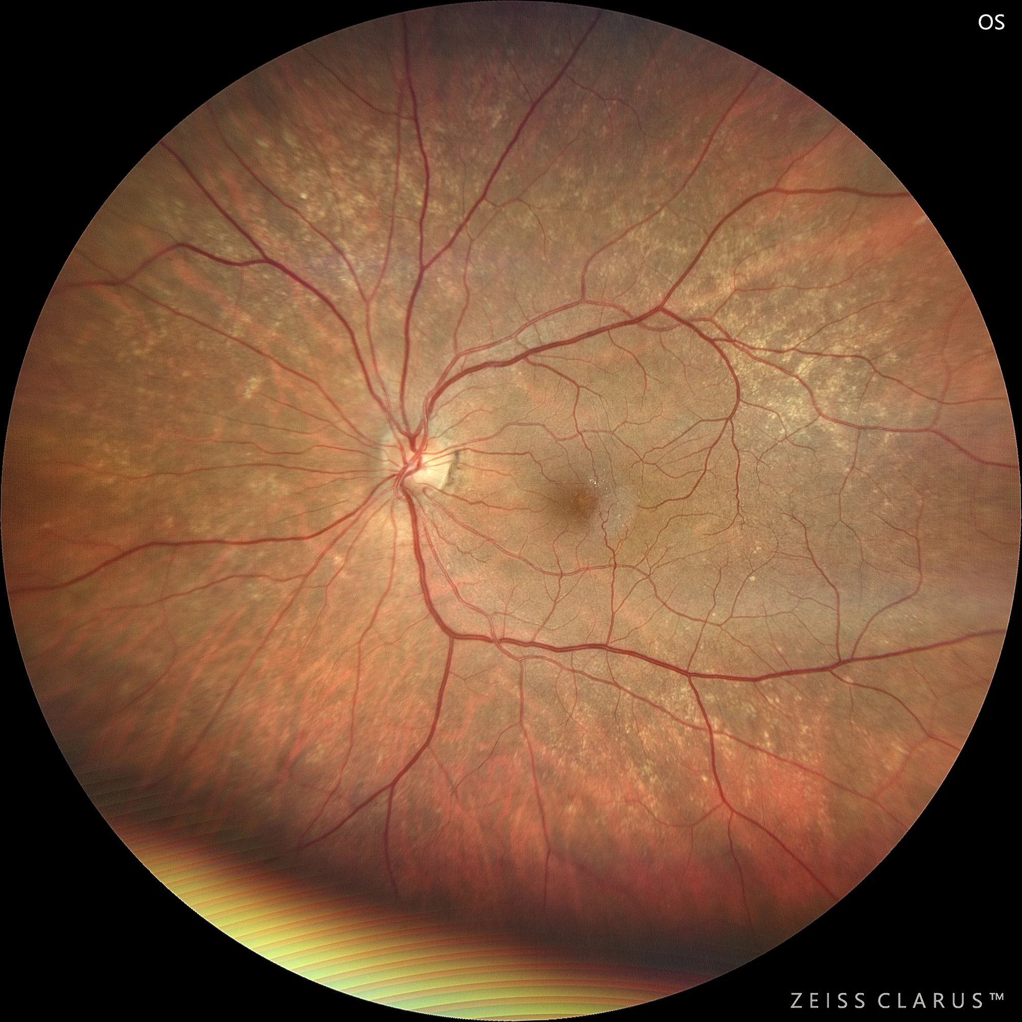
Color retinography showing a very characteristic image of this entity, a patch temporal to the macula with a shine, whitening, with microcysts, and telangiectatic-type vascular alterations.
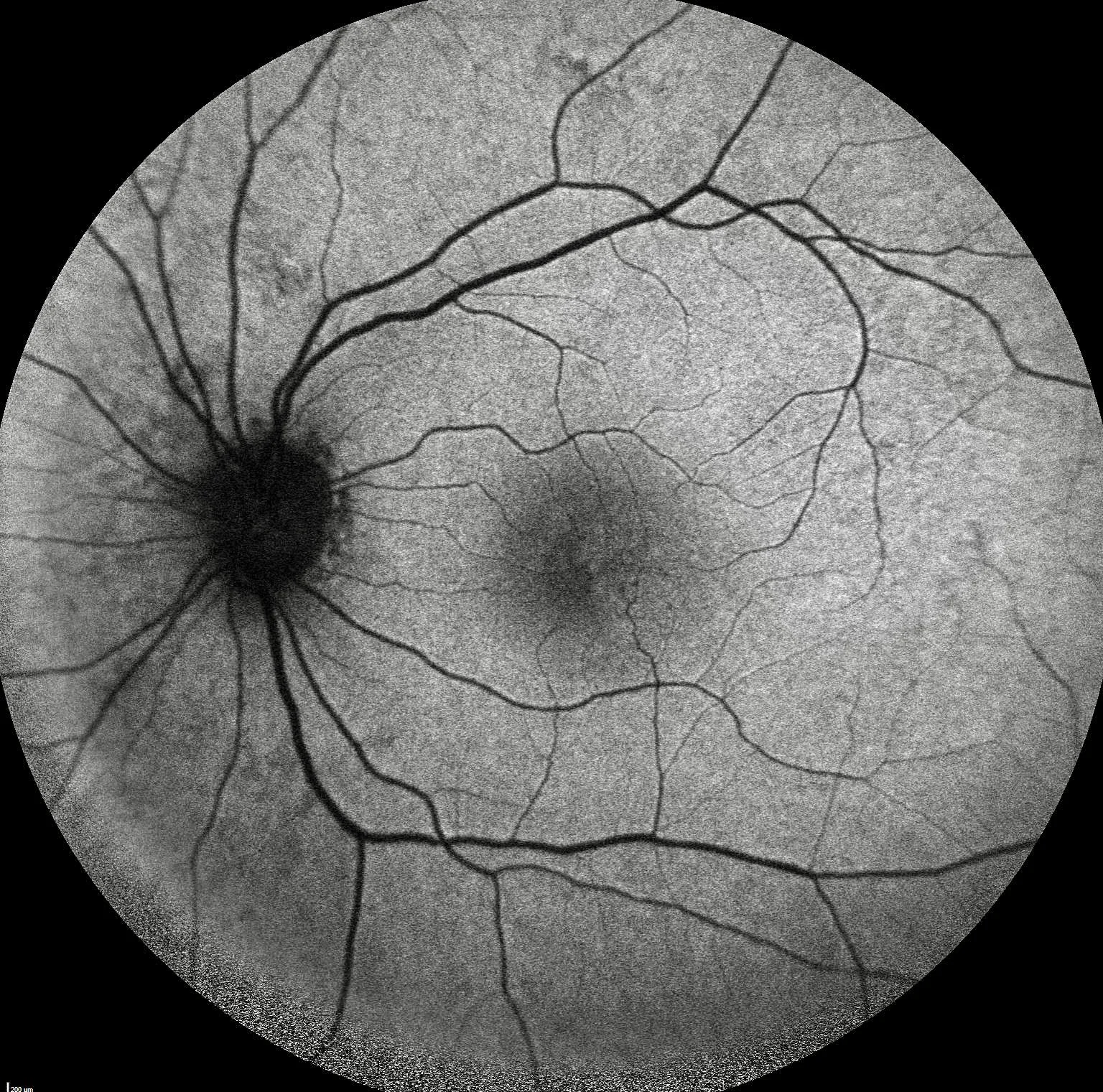
Loss of physiological hypoautofluorescence in the temporal half of the macula
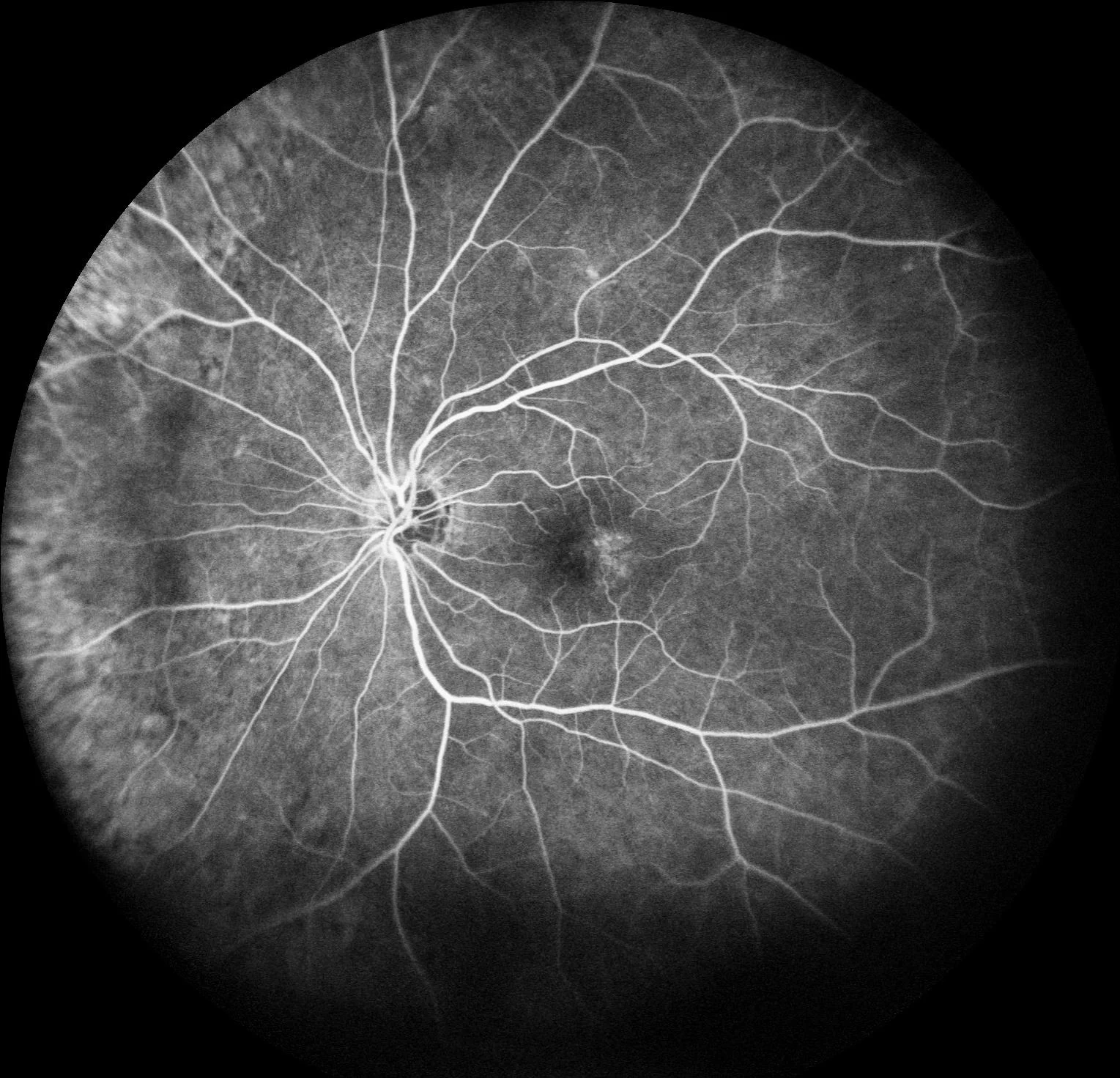
Angiography shows vascular redirection towards the temporal raphe of the macula, generating temporary vascular congestion with exudation at later stages.
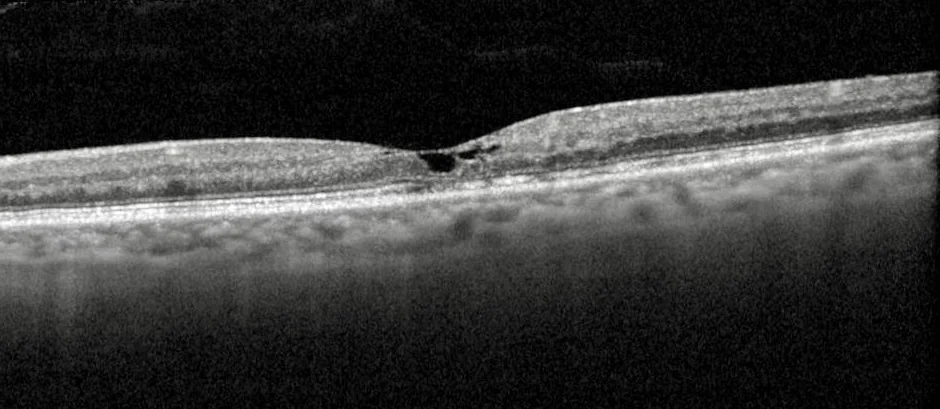
OCT is usually the most cost-effective test for diagnosing this condition. It shows intraretinal cavitations (not edema) in the temporal raphe of the macula of both eyes, making it almost pathognomonic.
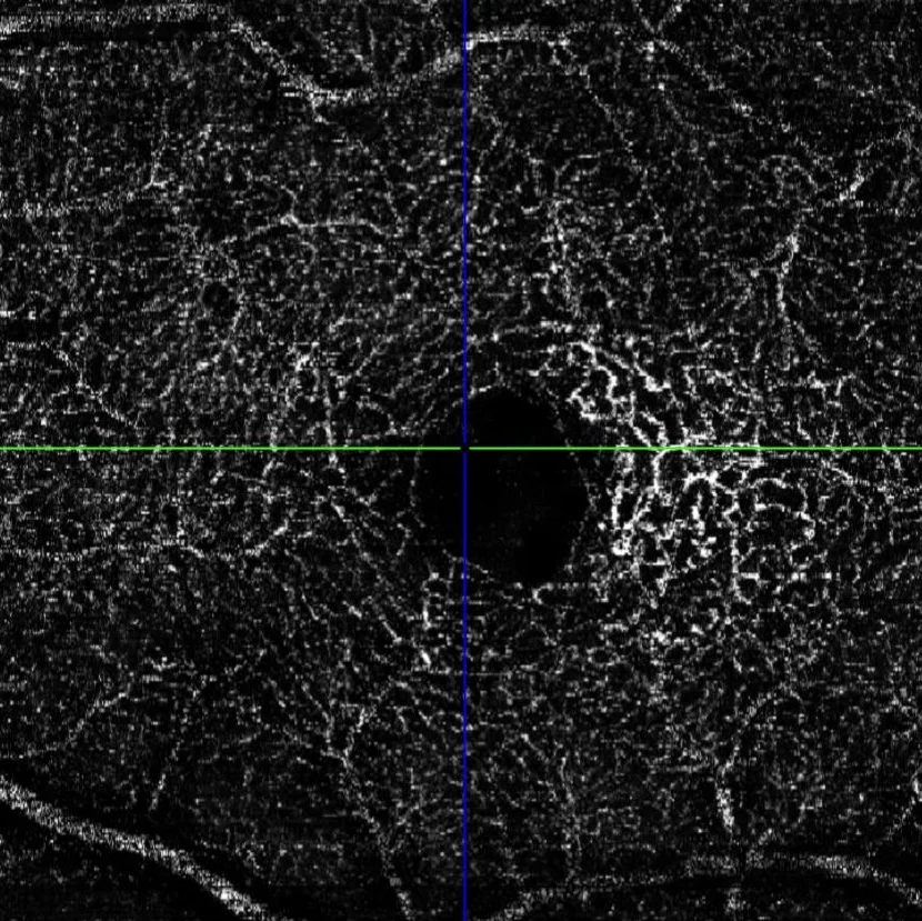
OCT angiography shows a prominent presence in the intermediate and deep plexus.
Description
Macular telangiectasias type 2, also known as idiopathic macular telangiectasias type 2 (MacTel 2), is a bilateral degenerative condition of the macula, which mainly affects middle-aged people. This pathology is characterized by dilation of perifoveal capillaries and abnormal proliferation of blood vessels in the retina, which can lead to progressive loss of central vision. Patients with MacTel type 2 usually present blurred vision and metamorphopsia. On clinical examination, retinal telangiectasias, crystalline deposits and cystic cavities in the inner retina can be seen. Optical coherence tomography (OCT) and fluorescein angiography are key tools for the diagnosis and monitoring of this disease, allowing detailed visualization of vascular and structural changes in the macula.