Moderate diabetic retinopathy
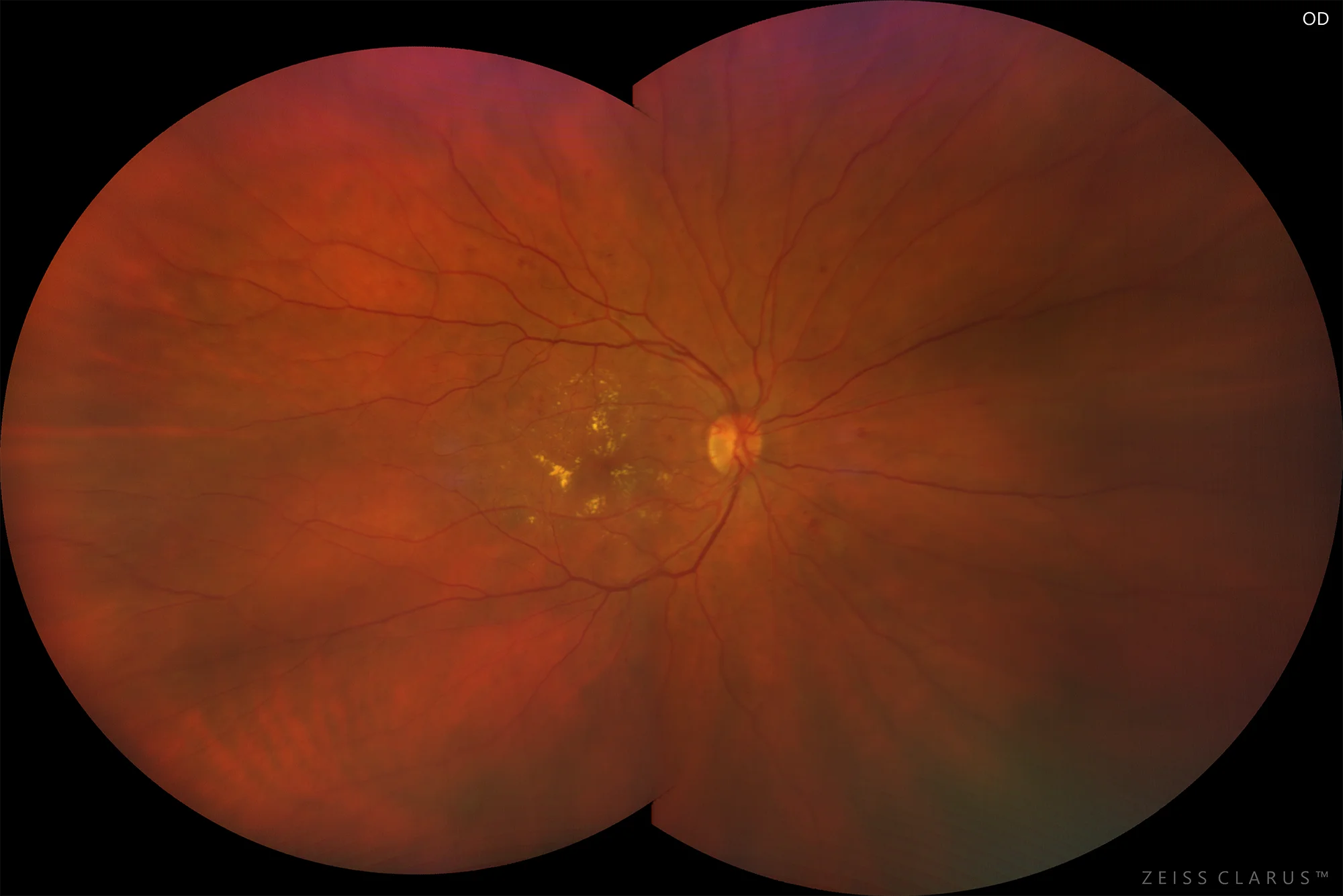
Figure 1. Retinography of the right eye showing the presence of hard exudates in the posterior pole and intraretinal hemorrhages in all four quadrants.
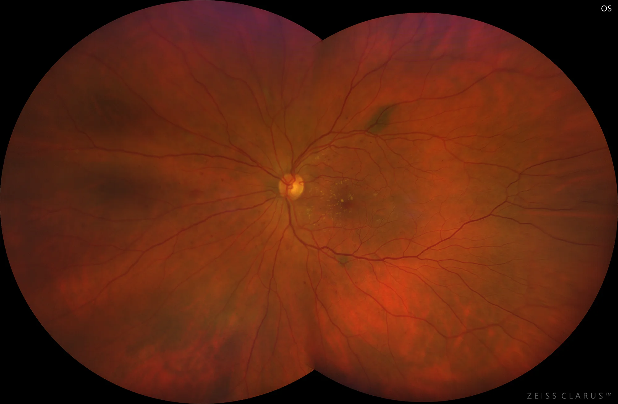
Figure 2. Retinography of the left eye showing the presence of hard perifoveal exudates and in the papillomacular bundle, as well as intraretinal hemorrhages in the four quadrants. Flat choroidal nevus at the end of the upper temporal arch.
Description
Diabetic retinopathy (DR) is, together with nephropathy and neuropathy, one of the microvascular complications of diabetes mellitus (DM), and is the main cause of irreversible visual loss in industrialized countries, especially in middle age. The duration of diabetes, the type of diabetes and metabolic control are the factors most directly related to the progression of DR. The appearance of microalbuminuria as a marker of microvascular disease increases the probability of developing DR.
Prevention of blindness due to DR involves regular eye examinations, the frequency of which will be determined by the type of diabetes and the state of the retina, among other factors.