Eales disease
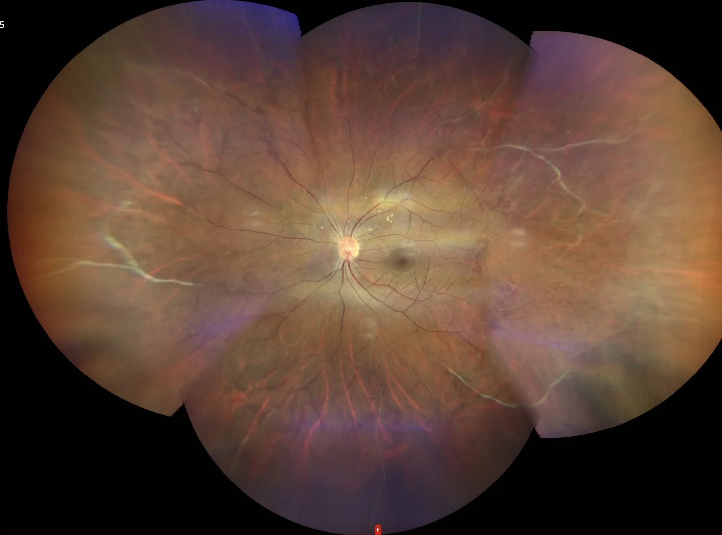
Color: montage of color retinography in which multiple vascular anomalies can be observed. We highlight the significant occlusive phlebitis and the bloodless image at its distal ends.
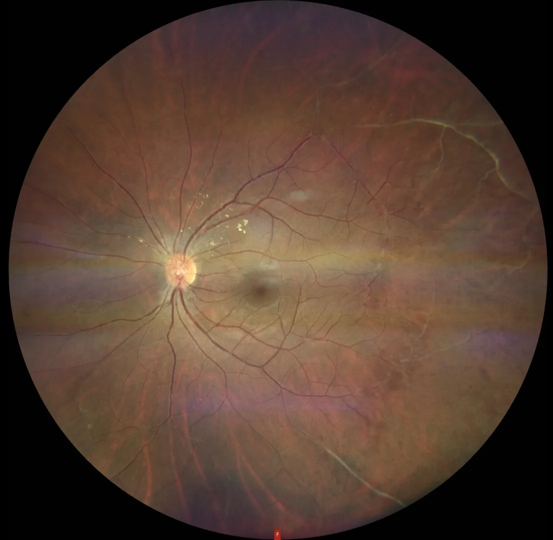
Color: Enlarged color image in the posterior pole in which vasculitis at the expense of the venous tree again stands out.
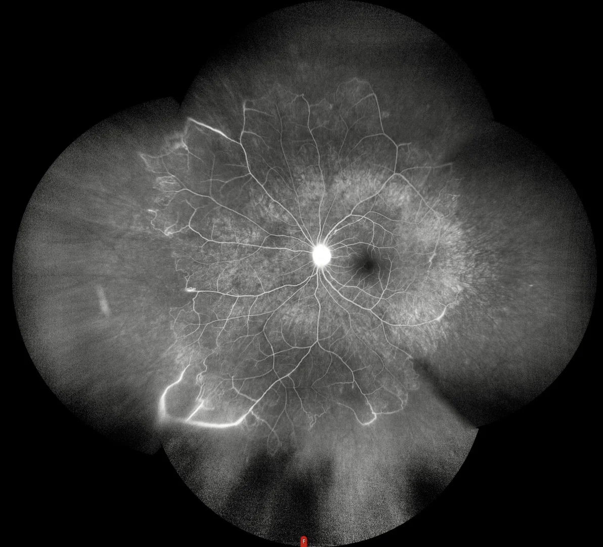
Fluorescein angiography: highlights the presence of vascular amputations in the mid-periphery, intraretinal microvascular abnormalities, microaneurysm-like hyperfluorescent points, and neovessels with fluorescein staining/diffusion.
Description
Eales disease is an idiopathic occlusive peripheral periphlebitis that primarily affects young men, particularly in the Indian subcontinent. It is characterized by inflammation, venous occlusion, and neovascularization, which may lead to recurrent vitreous hemorrhages and significant visual impairment. The pathogenesis is thought to involve a hypersensitivity to tuberculoprotein, supported by the presence of mycobacterial DNA in vitreous samples from affected individuals. Clinically, it manifests as peripheral retinal perivasculitis, often visible as a venous sheath with or without associated intraretinal hemorrhages. The disease progresses through stages of inflammation, occlusion, and neovascularization, with the potential for tractional retinal detachments if left untreated.