Retinal arterial macroaneurysm
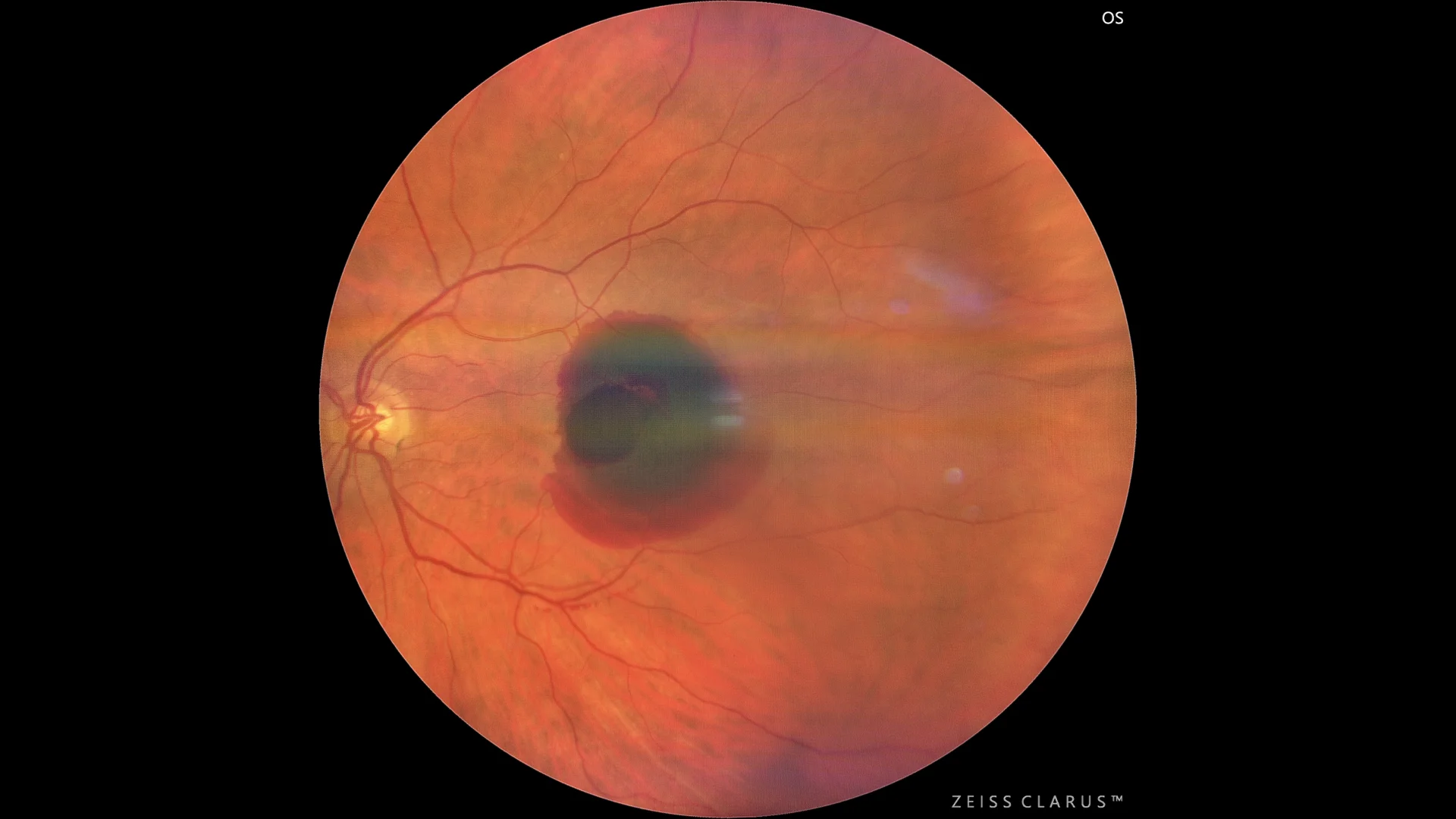
Color retinography showing retinal hemorrhage, with a subretinal component, with the retinal vessels above, and an internal sublimiting component.
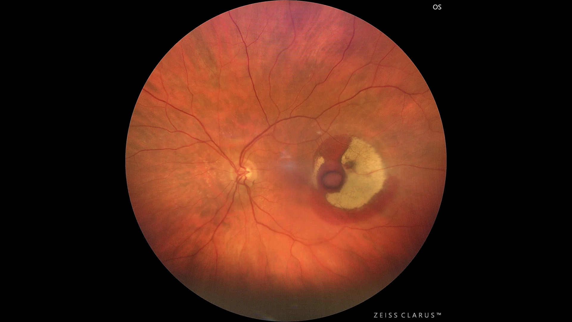
Color retinography image weeks later showing some reabsorption of the hemorrhage, with fibrinoid hematoma degradation, and the remnant of the sublimiting hematoma. In addition, the MAR can be seen in the center of the hemorrhage.
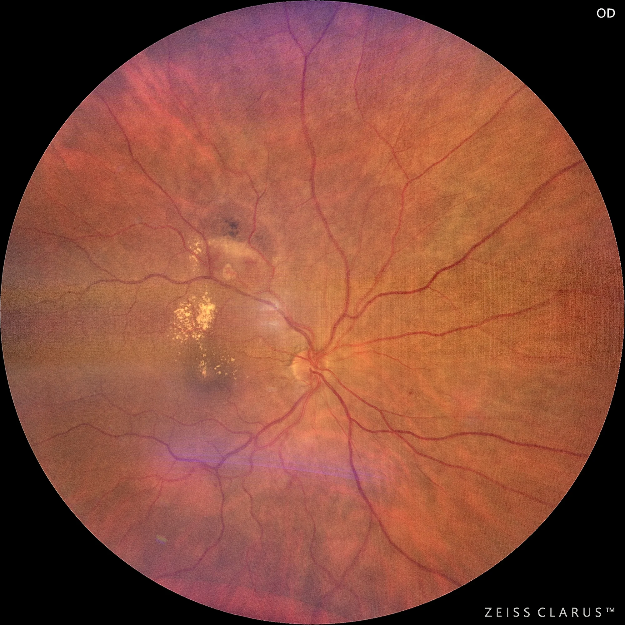
Color retinography of another MAR with exudative behavior, on the upper temporal arch of the RE, with hemorrhage in resolution, and with lipid material threatening the macula
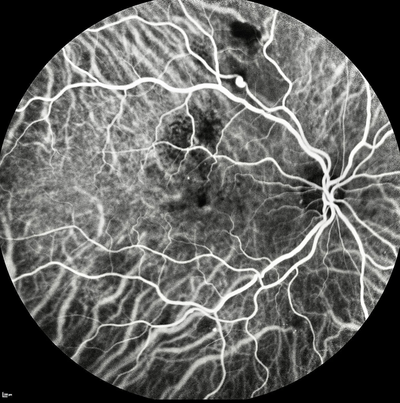
We use indocyanine green to correctly visualize the MAR without causing diffusion, which facilitates correct laser planning.
Description
Retinal arterial macroaneurysm (MAR) is an abnormal dilation of the retinal arteries, most common in older women and often associated with hypertension. On fundusography, MAR appears as a round, prominent lesion in the artery, which may be saccular or fusiform. It can cause retinal hemorrhages if it ruptures and lipid exudates from plasma leakage. Treatment varies depending on the size, location, and complications such as hemorrhages or macular edema.