Central retinal artery occlusion
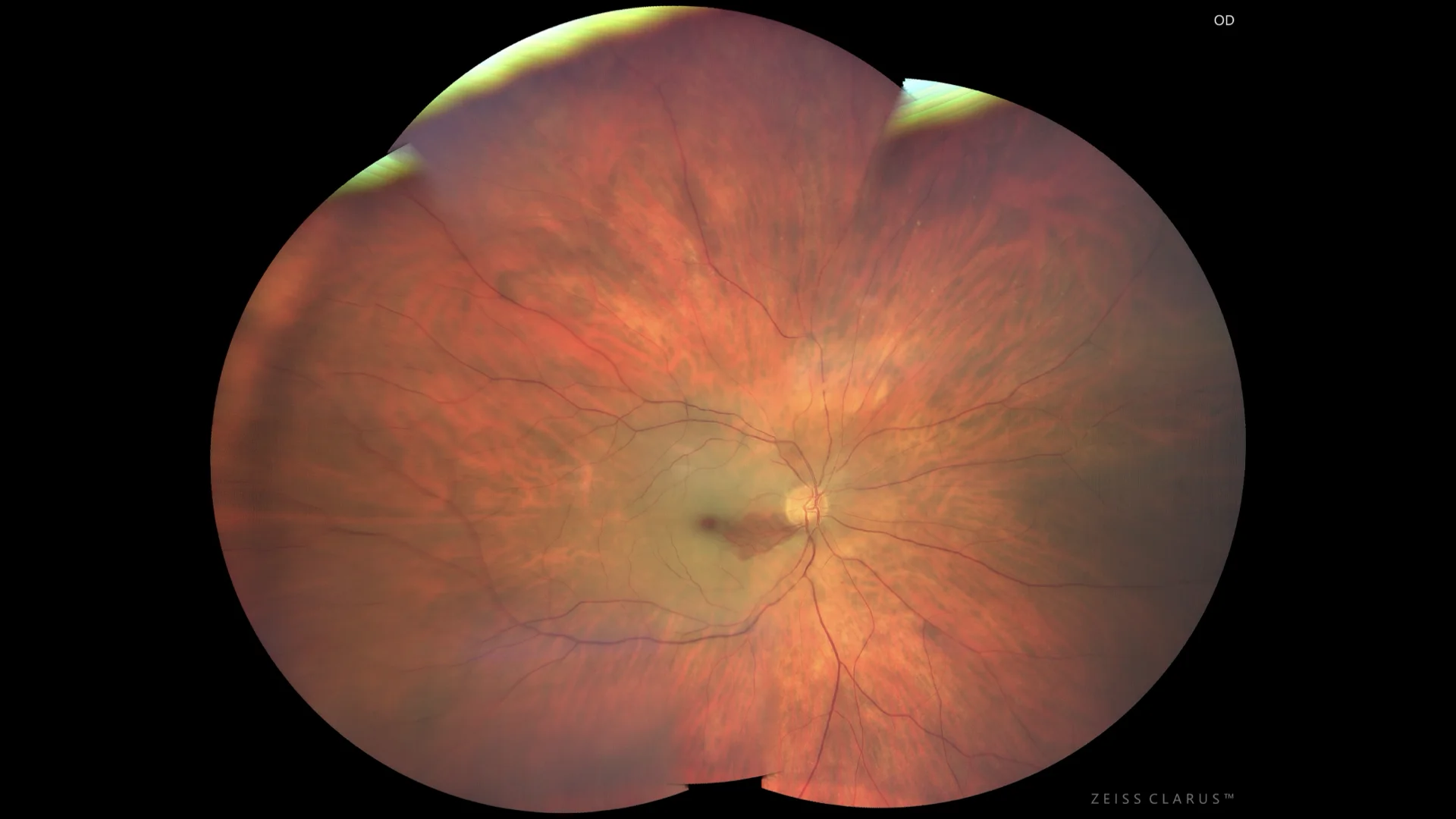
Color retinography showing a yellowish-white pallor in the posterior pole with a remnant in the papillo-macular bundle that remains permeable due to the existence of a cilioretinal artery in this case.
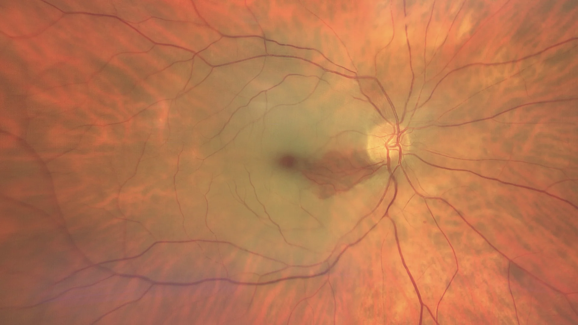
Enhanced color retinography in the posterior pole showing arterial and venous vascular segmentation, characteristic of this type of condition.
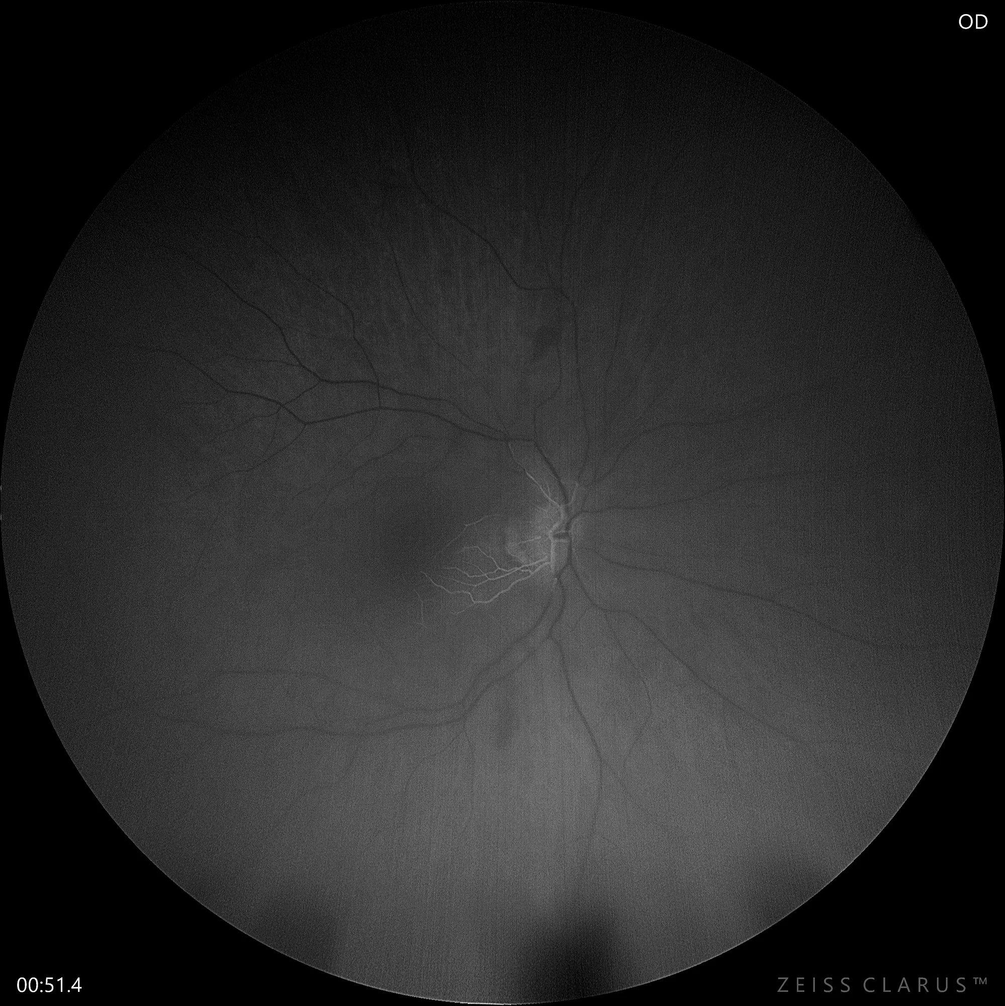
Fluorescein angiography in which we observe the filling of only the cilioretinal artery as well as the venous return depending on this arterial branch.
Description
Central retinal artery occlusion (CRAO) is an ophthalmic emergency usually caused by an embolus blocking the central retinal artery, leading to sudden vision loss. On fundusography, it is characterized by diffuse retinal pallor with a reddened macula, known as the cherry red spot sign. Other findings may include narrowing of arterioles and segmentation of the blood column. Urgent treatment aims to restore blood flow and may include maneuvers to decrease intraocular pressure, administration of agents that reduce blood viscosity, and therapies to dissolve or dislodge the embolus. Prompt detection and treatment are critical to attempting to restore visual function.