Chronic inferior branch retinal artery occlusion
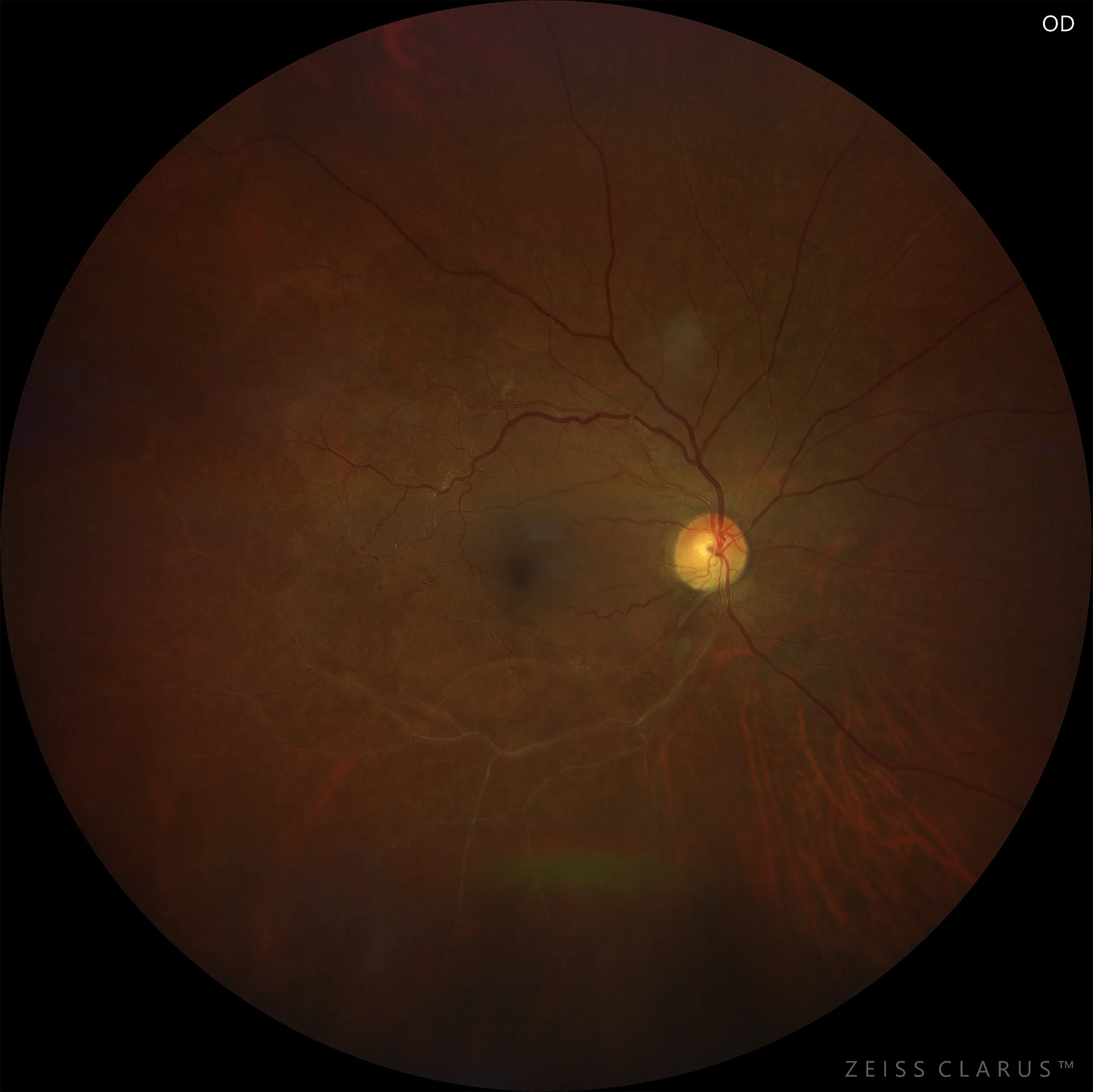
Figure 1. Color retinography showing hyalinization of the inferior temporal vessels of the right eye, indicating chronic arterial occlusion.
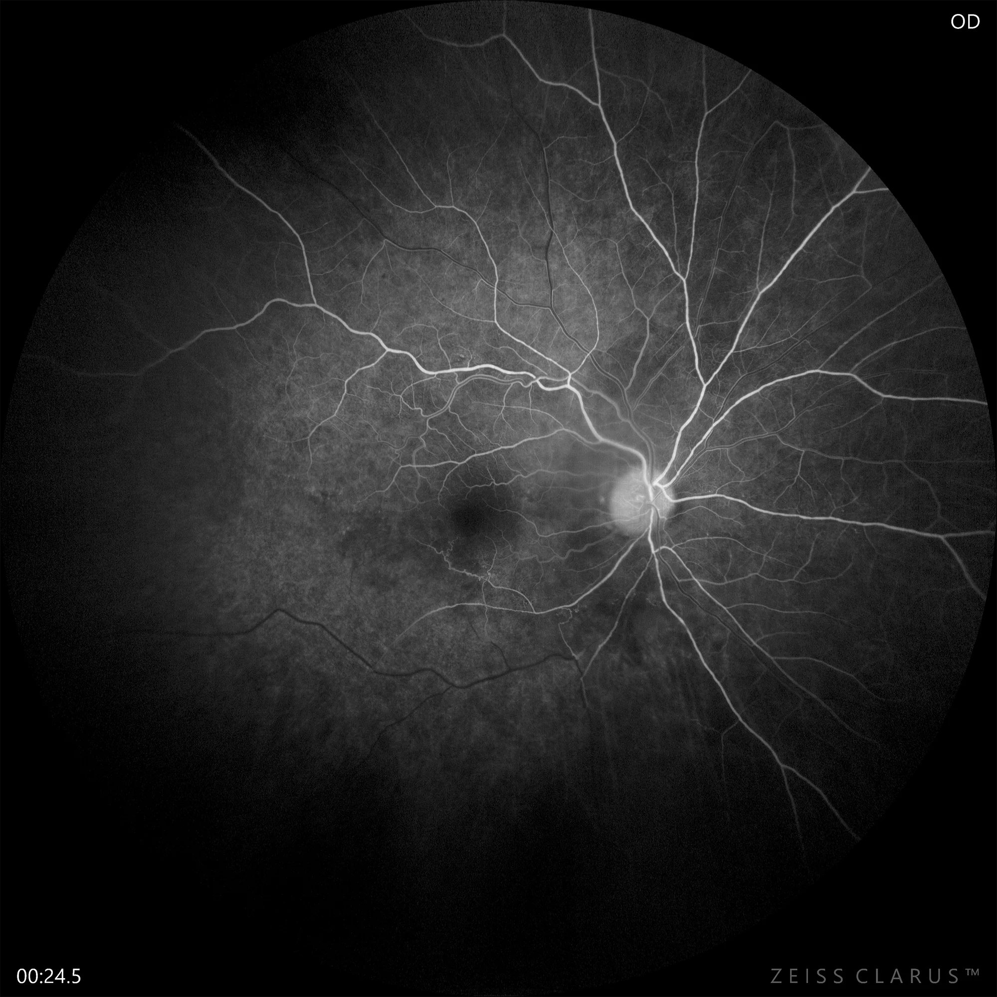
Figure 2. Fluorescein angiography of the right eye showing the inferior temporal arterial occlusion and the presence of collaterals at different times of the venous phase: 24 seconds
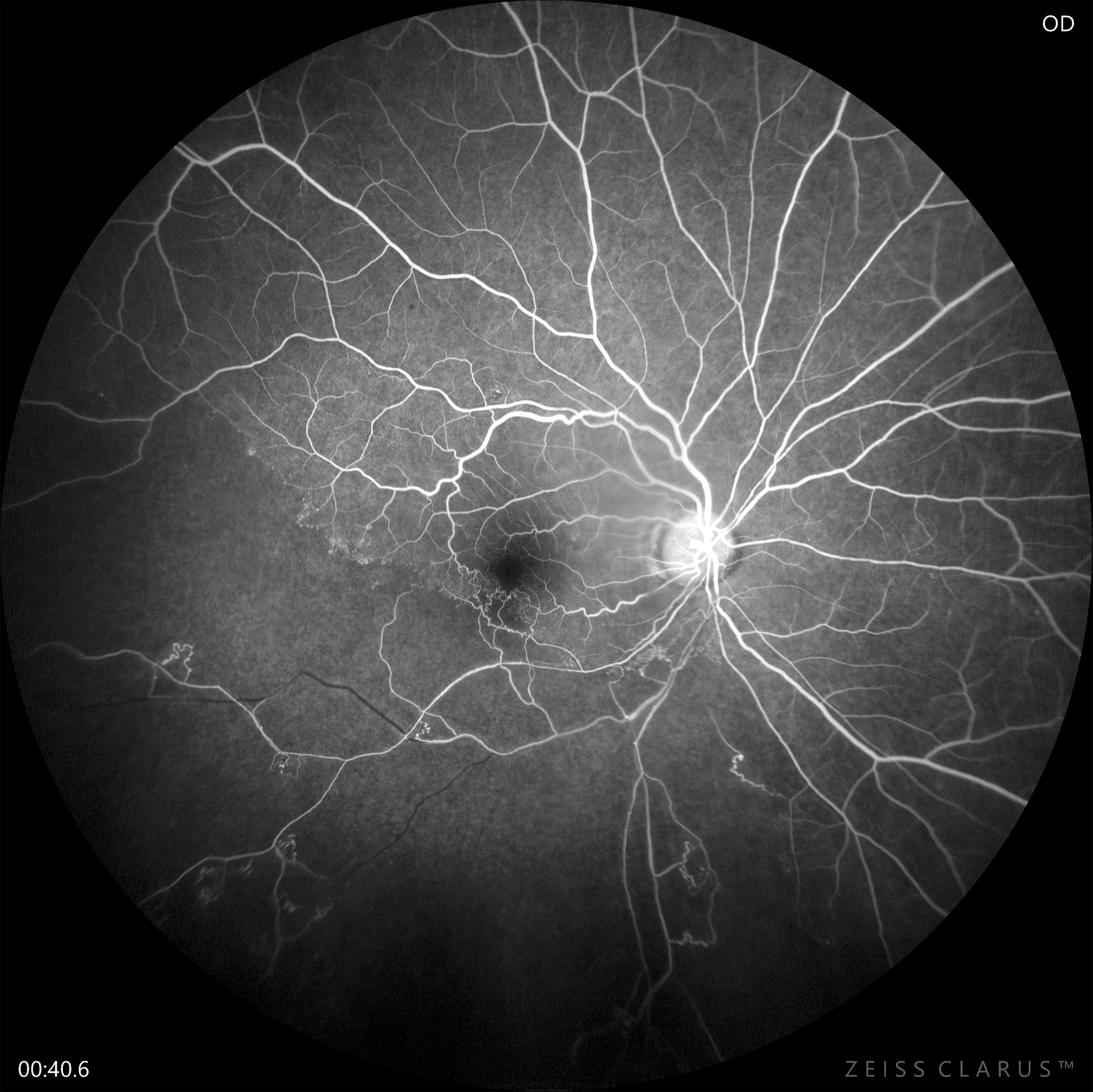
40 seconds
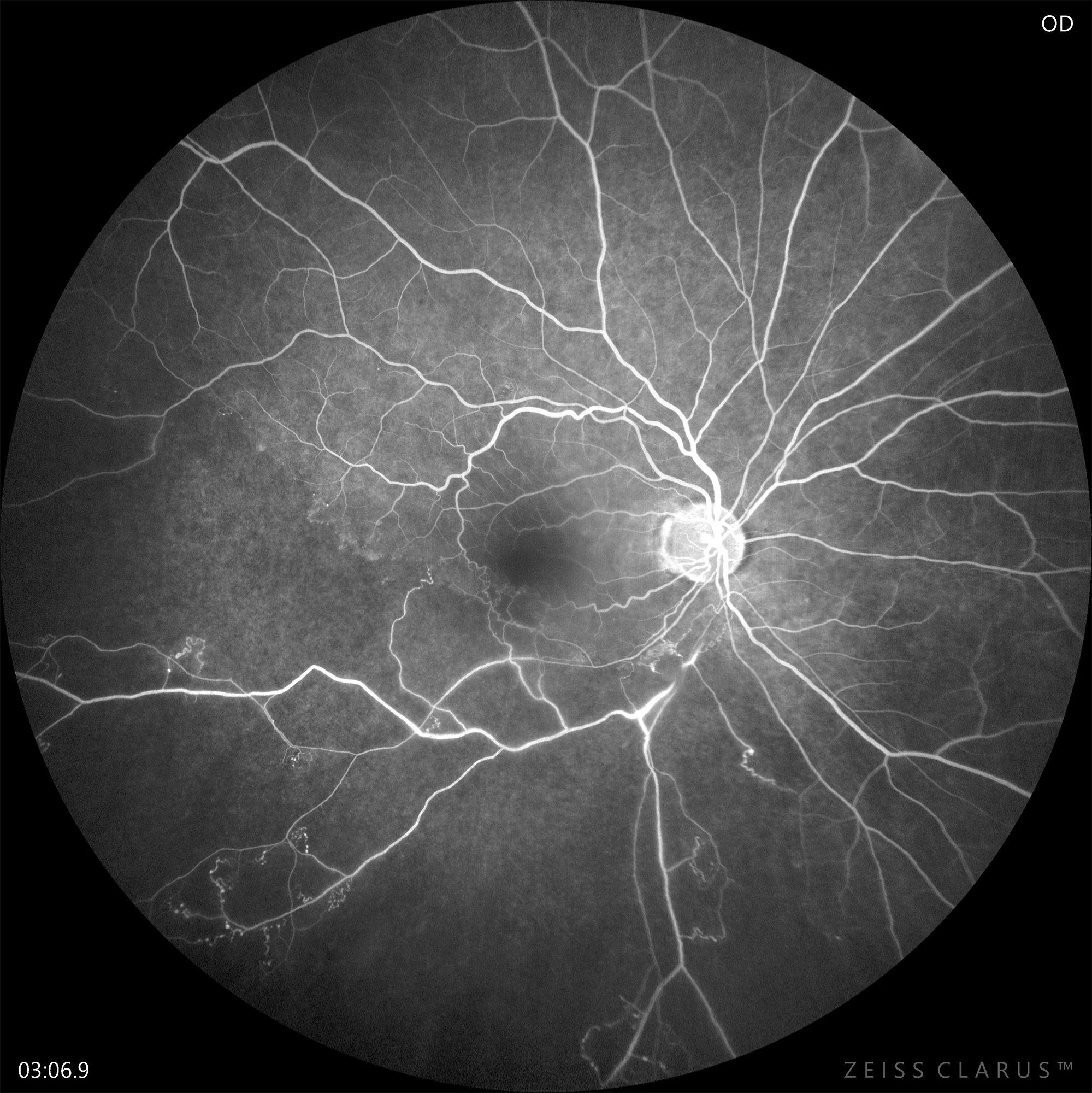
3 minutes
Description
Symptomatic retinal arterial occlusion is an ophthalmologic emergency that requires immediate evaluation. It is an obstruction of blood flow that can be caused by an embolus, vasculitis, trauma to the vessel wall, or a spasm. Vision loss occurs in the area of retinal ischemia. This pathology is associated with high cerebral and cardiovascular morbidity and mortality. Some patients present silent and simultaneous ischemic strokes, which is why it is recommended to perform a blood test with a complete blood count, platelet count, coagulation, ESR, C-reactive protein, and glycosylated hemoglobin; an electrocardiogram and an echocardiogram if necessary; a cranial magnetic resonance imaging or CT scan; and a Doppler ultrasound of the supra-aortic trunks or magnetic resonance angiography or CT angiography of the carotid arteries.