< back
Congenital simple hamartoma of RPE

Color retinography (Clarus 700, Zeiss, detail): Small brownish-black pigmented lesion in the macular area.
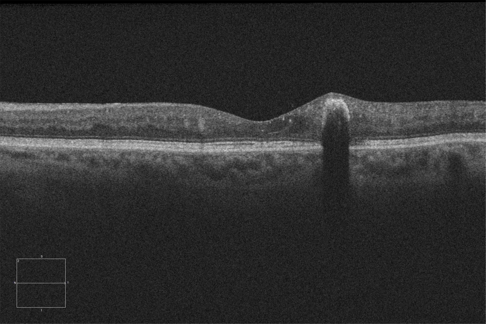
Optical coherence tomography (Cirrus-HD 5000, Zeiss) Protruding hyperreflective lesion in the inner retina with abrupt margins and a posterior hyporeflective acoustic shadow.
Description
Simple hamartoma of the retinal pigment epithelium is a highly pigmented, flat, solitary lesion of a congenital and benign nature, very rare.
OCT shows a characteristic pattern, which together with fundus imaging techniques allows differentiation from other more common fundus lesions. The visual prognosis is usually good, especially in cases with extrafoveal location.