Lacquer Streak
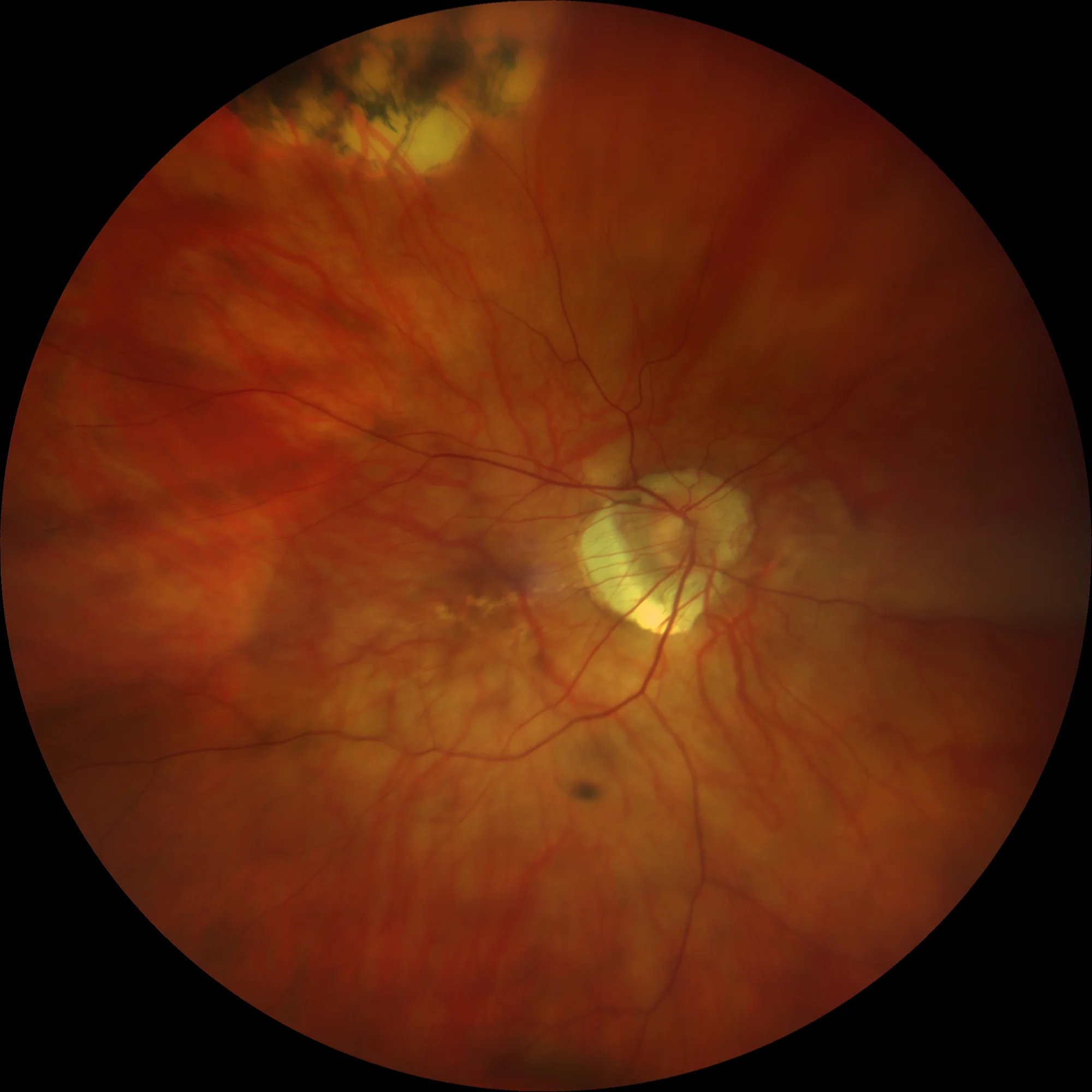
Retinography OD: Linear yellowish-white lesion that corresponds to a lacquer streak that crosses the macula from the papilla to the temporal sector.
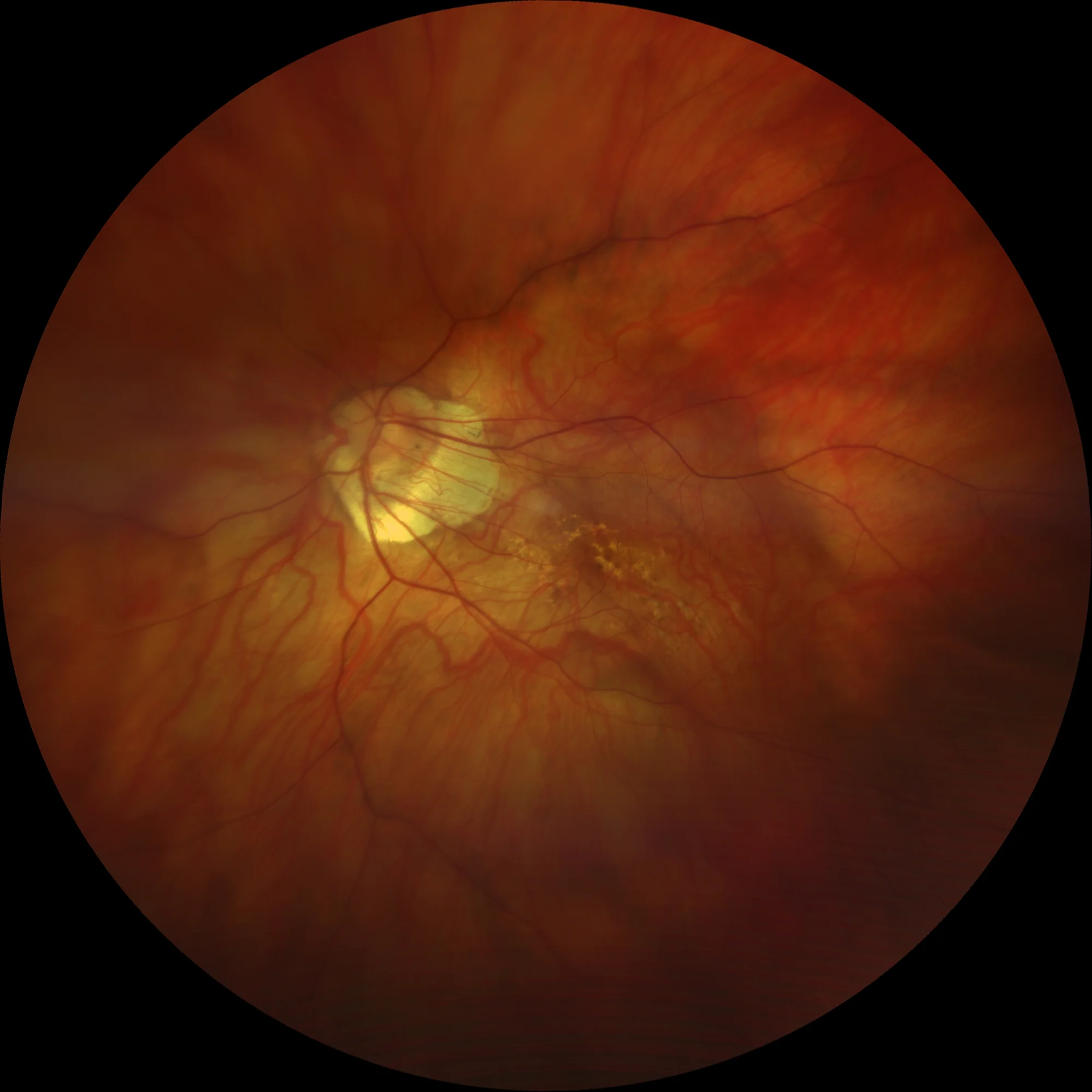
Left retina: Branched lacquer streak in macular area.
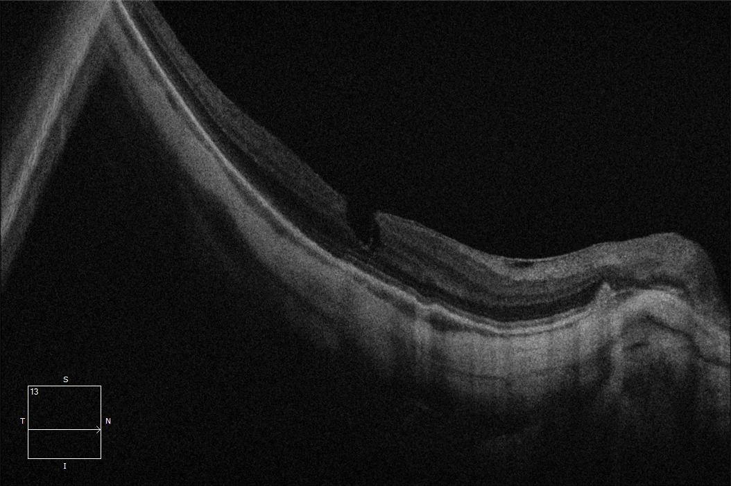
Macular OCT OD: Posterior staphyloma. Presence of epiretinal membrane with pseudohole image. Irregularity of the pigmented epithelium that could correspond to lacquer streak.
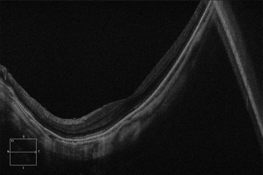
Macular OCT LE: Posterior staphyloma with preserved foveal profile. No structural alterations related to the lacquer streak are observed in the OCT, which may be common.
Description
Lacquer striae are considered to be healed mechanical tears of the choriocapillaris-Bruch’s membrane complex. They appear as single or multiple, irregular, yellowish-white linear lesions in or around the macular area. Lacquer striae are included within the neovascular myopic maculopathy of the ATN classification as the first stage, since they are a predisposing factor for the development of neovascular membranes in highly myopic individuals. The formation of lacquer striae appears to be related to axial elongation and posterior staphyloma. The same mechanism that produces choroidal thinning produces thinning of Bruch’s membrane leading to its fragmentation and rupture, producing lacquer striae. In response to these tears, the formation of choroidal neovascularization can be triggered through the tear, suggesting that these membranes correspond to an abnormal response of the healing mechanism. However, once the tear is covered by scar tissue, a membrane rarely appears. Another complication that these streaks can cause is their progression to the formation of chorioretinal atrophy plaques. Occasionally, when a lacquer streak occurs, subretinal bleeding or hemorrhage (vanishing hemorrhage) may occur with mild visual loss. It is important to differentiate these hemorrhages from those secondary to myopic neovascular membranes (NVM), since the evanescent hemorrhages of myopes do not require treatment and are usually reabsorbed on their own with recovery of visual acuity.