Lacquer Streak
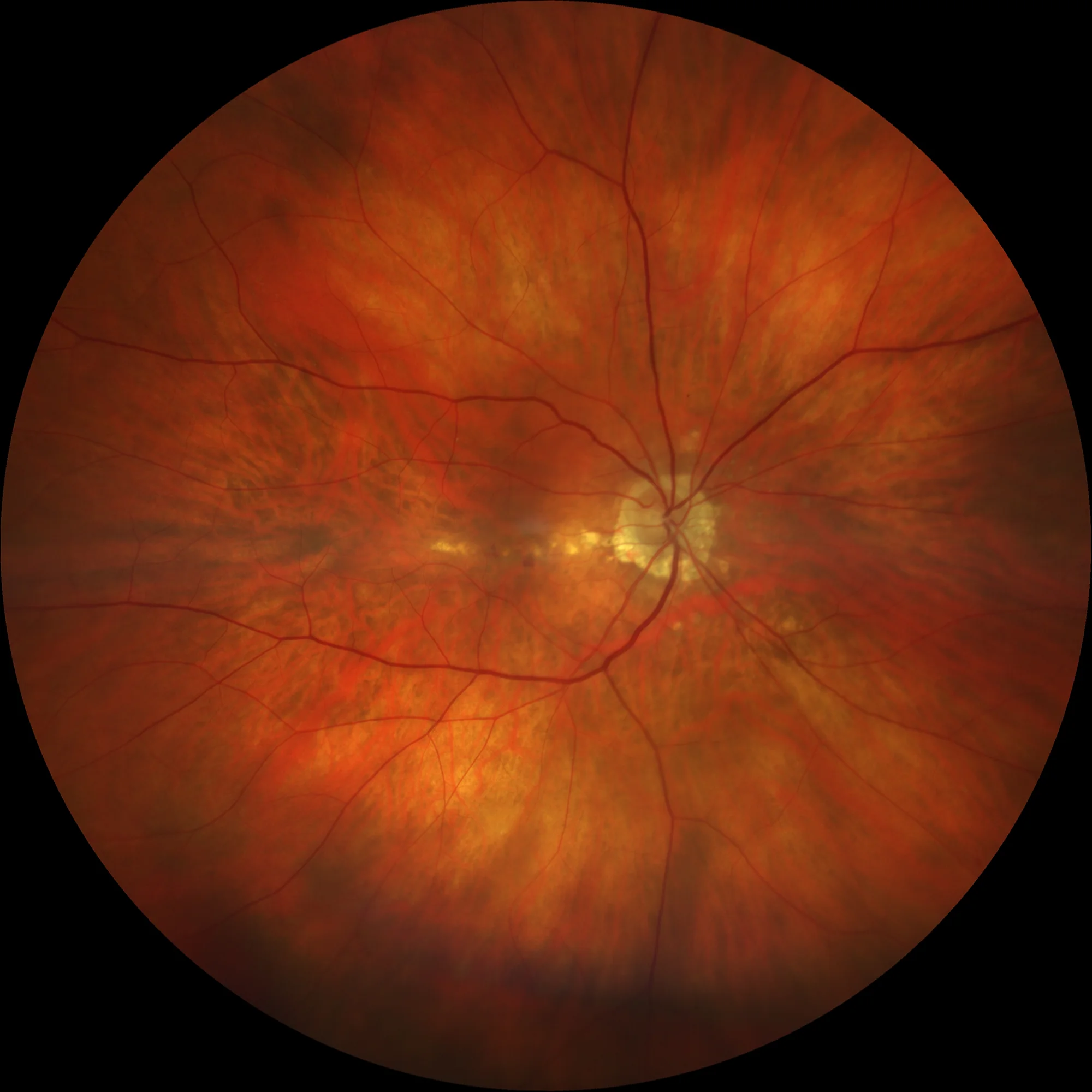
Retinography of the OD prior to treatment with anti-VEGF injections (2022): Linear yellowish-white lesion corresponding to a lacquer streak crossing the macula from the papilla to the temporal sector. Two small juxtafoveal microhemorrhages are observed.
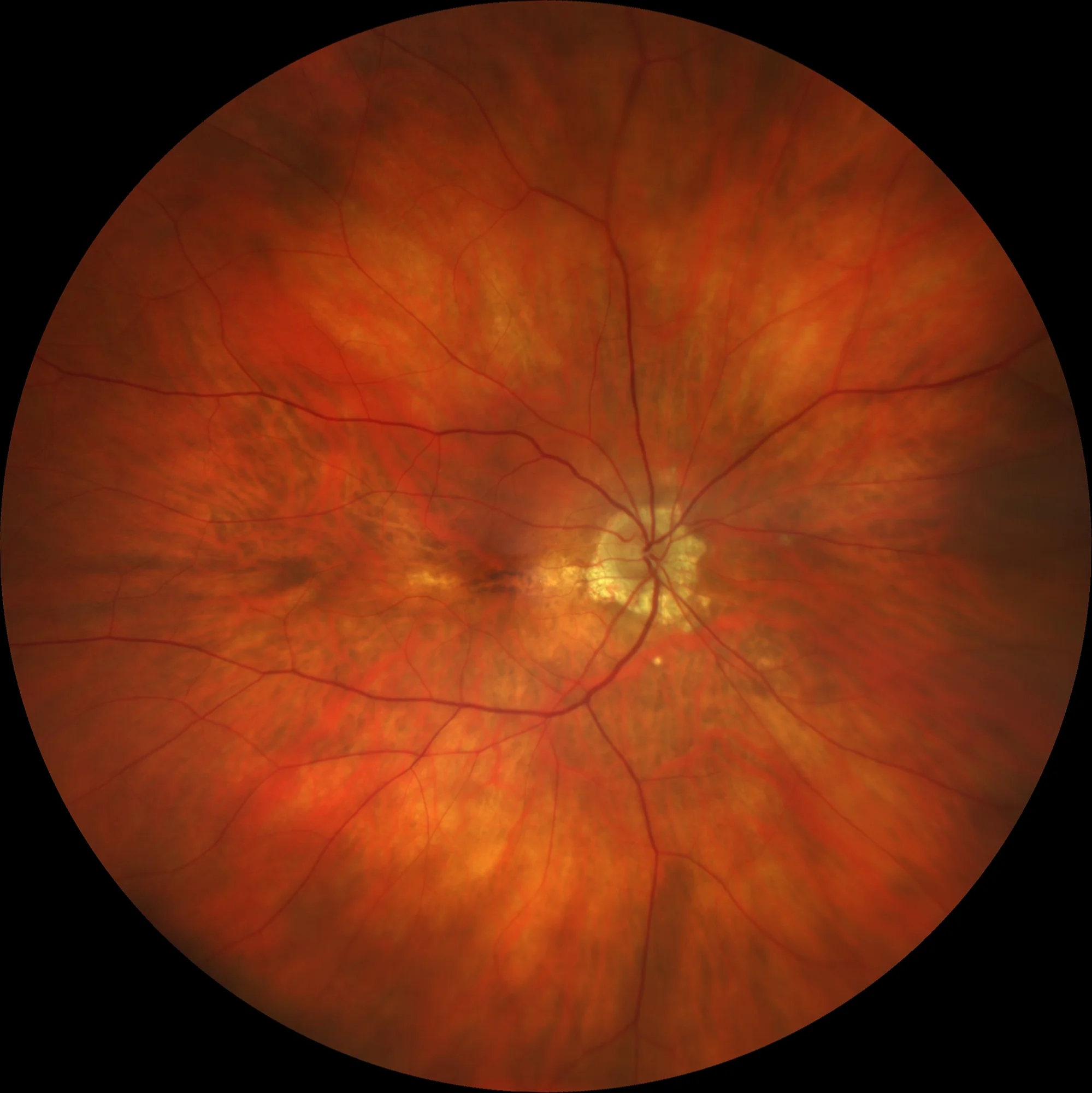
Retinography of the OD prior to treatment with anti-VEGF injections (2022): Linear yellowish-white lesion corresponding to a lacquer streak crossing the macula from the papilla to the temporal sector. Two small juxtafoveal microhemorrhages are observed.
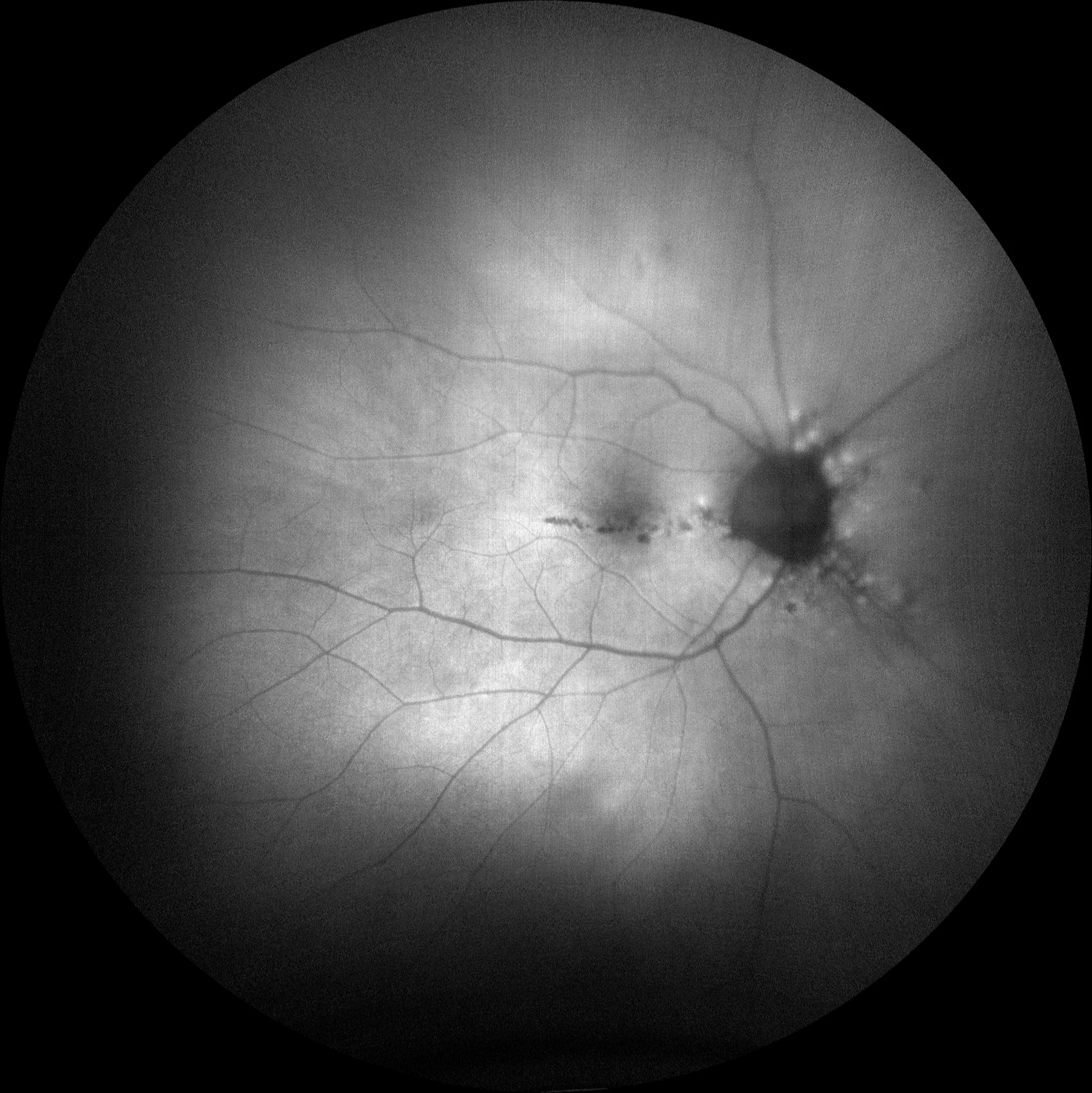
Pre-treatment autofluorescence OD (2022): The autofluorescence shows an alteration of the RPE along the path of the streak. Two small points of hypoautofluorescence can also be seen adjacent to the lacquer streak that correspond to the microhemorrhages by creating a screen effect.
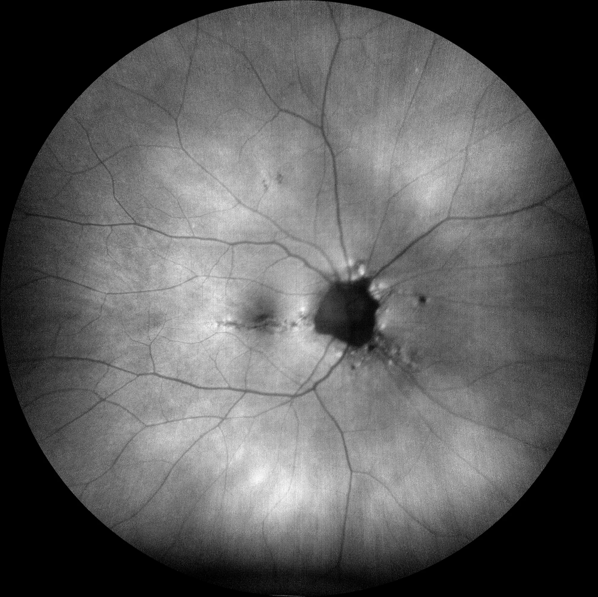
Post-treatment autofluorescence OD (2024): It shows a slight increase in the pigmentary alteration at the central level compared to the previous one.

OCT macular pre-treatment OD (2022): The OCT shows central retinal thickening with a subretinal hyperreflective lesion and destructuring of the outer retina, due to the presence of an active MNV. A small intraretinal cyst is also observed.
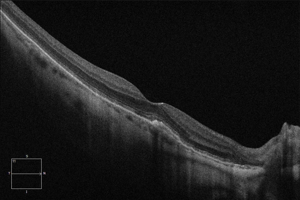
Post-treatment macular OCT OD (2024). We can observe a flat juxtafoveal retinal pigment epithelium detachment (PED), with a fibrovascular appearance and without associated fluid, which corresponds to the presence of a currently inactive neovascular membrane.
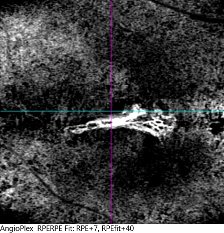
Macular angio-OCT OD post-treatment. The presence of choroidal neovascularization is observed below the DEP, in the path of the Lacquer stria.
Description
Lacquer striae are considered to be healed mechanical tears of the choriocapillaris-Bruch’s membrane complex. They appear as single or multiple, irregular, yellowish-white linear lesions in or around the macular area. Lacquer striae are included within the neovascular myopic maculopathy of the ATN classification as the first stage, since they are a predisposing factor for the development of neovascular membranes in highly myopic individuals. The formation of lacquer striae appears to be related to axial elongation and posterior staphyloma. The same mechanism that produces choroidal thinning produces thinning of Bruch’s membrane leading to its fragmentation and rupture, producing lacquer striae. In response to these tears, the formation of choroidal neovascularization can be triggered through the tear, suggesting that these membranes correspond to an abnormal response of the healing mechanism. However, once the tear is covered by scar tissue, a membrane rarely appears. Another complication that these streaks can cause is their progression to the formation of chorioretinal atrophy plaques. Occasionally, when a lacquer streak occurs, subretinal bleeding or hemorrhage (vanishing hemorrhage) may occur with mild visual loss. It is important to differentiate these hemorrhages from those secondary to myopic neovascular membranes (NVM), since the evanescent hemorrhages of myopes do not require treatment and are usually reabsorbed on their own with recovery of visual acuity.