Retinal pigment epithelium tear
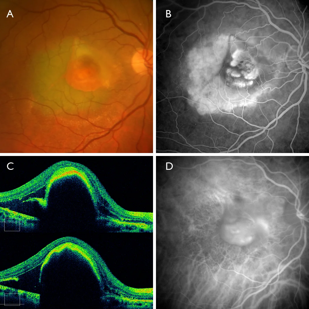
• 50º Retinography (FF450 IR plus, Zeiss): Color (A), Fluorescein angiography (B), Indocyanine green angiography (C): Neovascular membrane clearly visible on AFG with large RPE tear in the temporal area with hyperfluorescence due to window effect. The yellowish coloration of intravenous fluorescein can be seen in the color image. • Optical coherence tomography (Cirrus HD5000, Zeiss): Large RPE detachment with RPE tear clearly visible in two horizontal slices in the temporal area.
Description
Retinal pigment epithelial (RPE) tear is a serious complication of age-related macular degeneration, which can occur spontaneously or, more frequently, after antiangiogenic therapy. It is associated in most cases with large vascularized RPE detachments (PEDs) and usually entails a severe loss of visual acuity.