Tuberous sclerosis
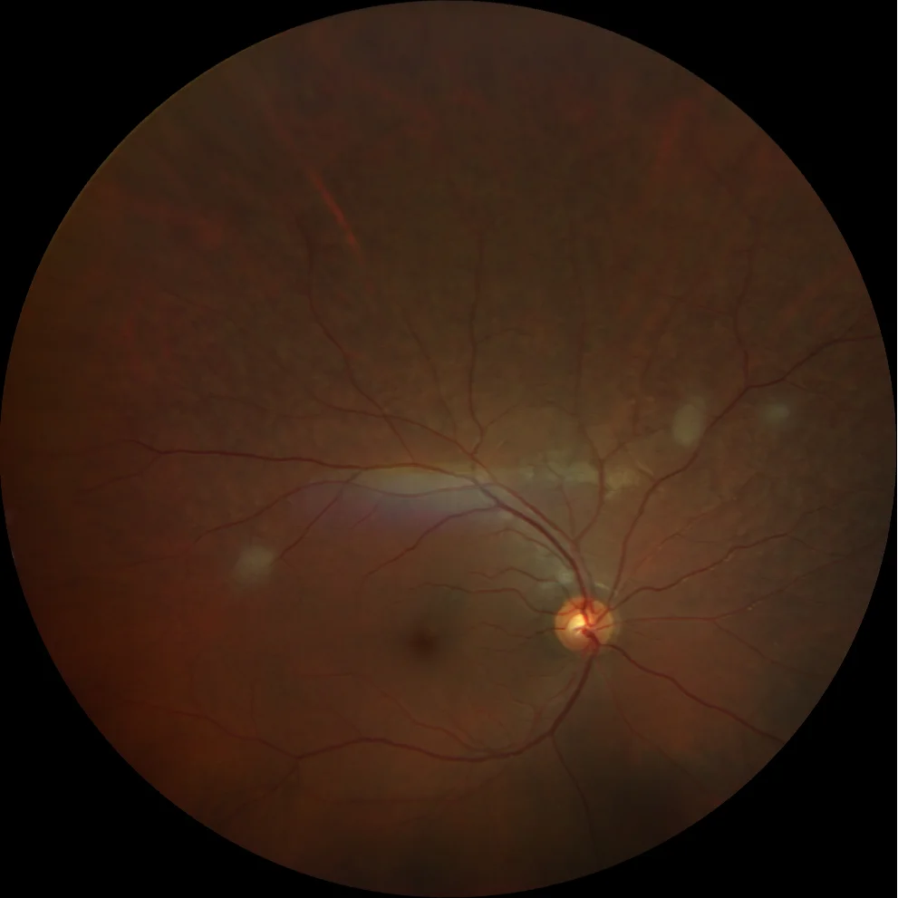
Color fundusography (Clarus 500, Carl Zeiss Meditec ASG, Jena, Germany) of the right and left eyes, respectively, showing astrocytic retinal hamartomas not involving the macula, as well as a superotemporal achromic path in the left eye.
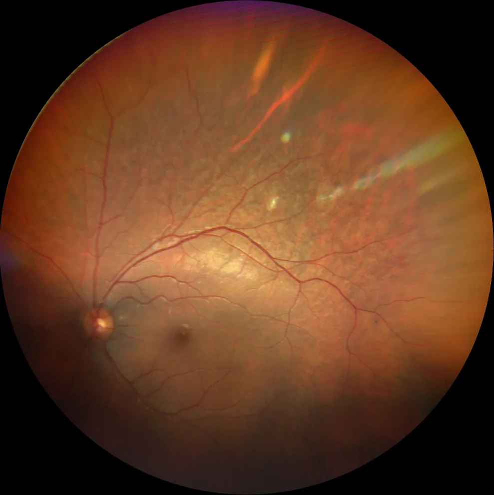
Color fundusography (Clarus 500, Carl Zeiss Meditec ASG, Jena, Germany) of the right and left eyes, respectively, showing astrocytic retinal hamartomas sparing the macula, as well as a superotemporal achromic path in the left eye.
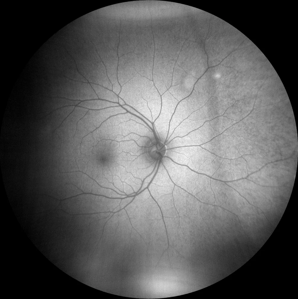
Autofluorescence image (Clarus 500, Carl Zeiss Meditec ASG, Jena, Germany) showing mild hyperautofluorescence in the lesions of the right eye, while in the left eye the lesions are hyperautofluorescent (compatible with calcification). The achromic patch appears hypoautofluorescent.
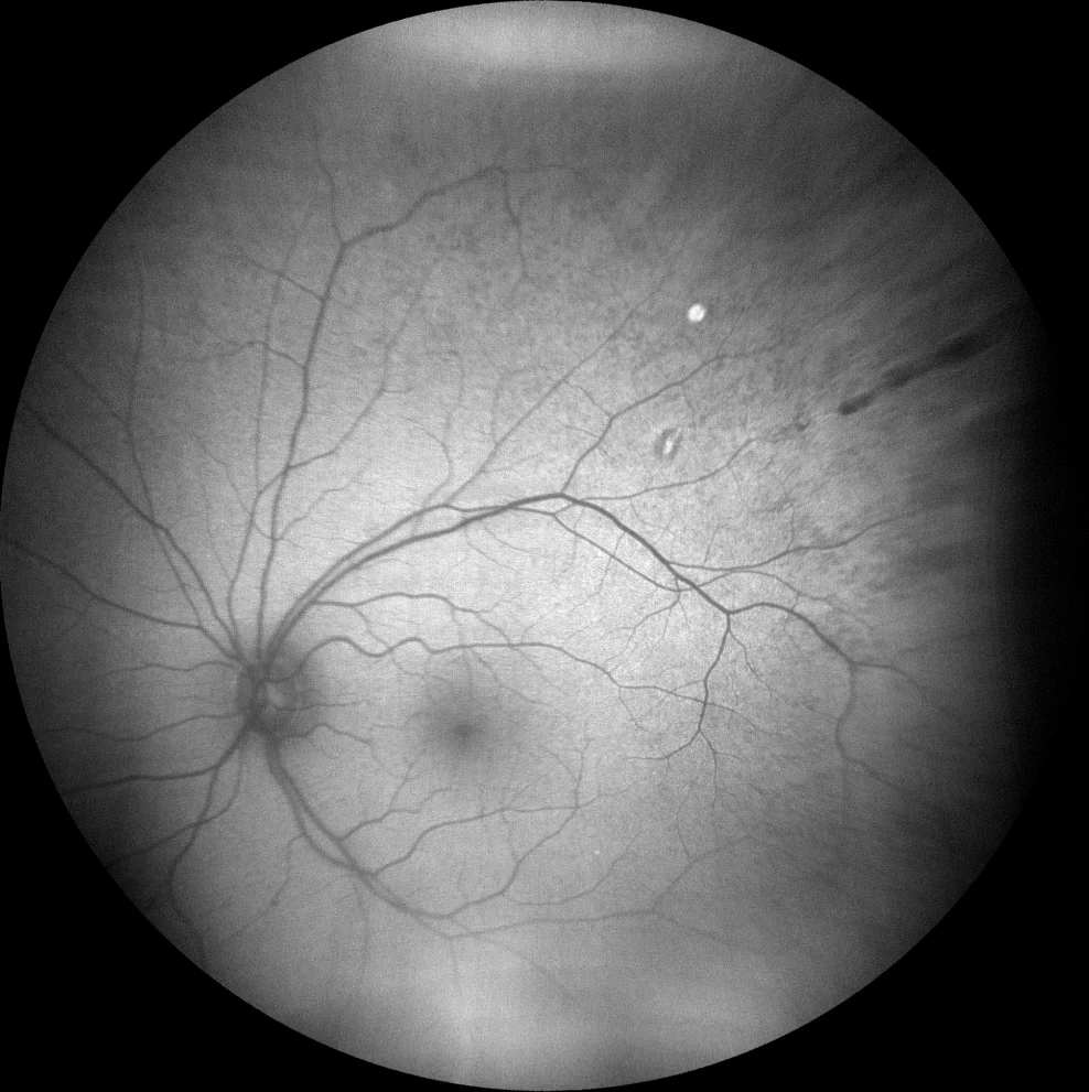
Autofluorescence image (Clarus 500, Carl Zeiss Meditec ASG, Jena, Germany) showing mild hyperautofluorescence in the lesions of the right eye, while in the left eye the lesions are hyperautofluorescent (compatible with calcification). The achromic patch appears hypoautofluorescent.
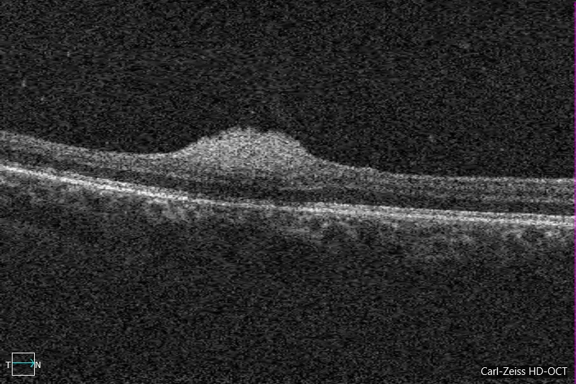
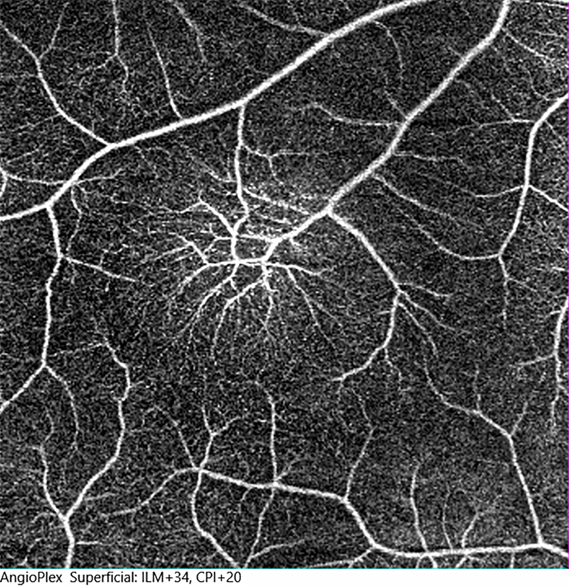
. Optical coherence tomography angiography (OCT-A) of the three retinal astrocytic hamartomas of the right eye. E and F: Horizontal B-scan over the temporal (E) and superonasal (F) hamartomas, showing segmentation for the superficial capillary plexus, where the lesions and their vasculature are located. G: OCT-A en face of the superficial plexus of the temporal retinal astrocytic hamartoma, showing a single feeding artery and a single draining vein, branching pattern.
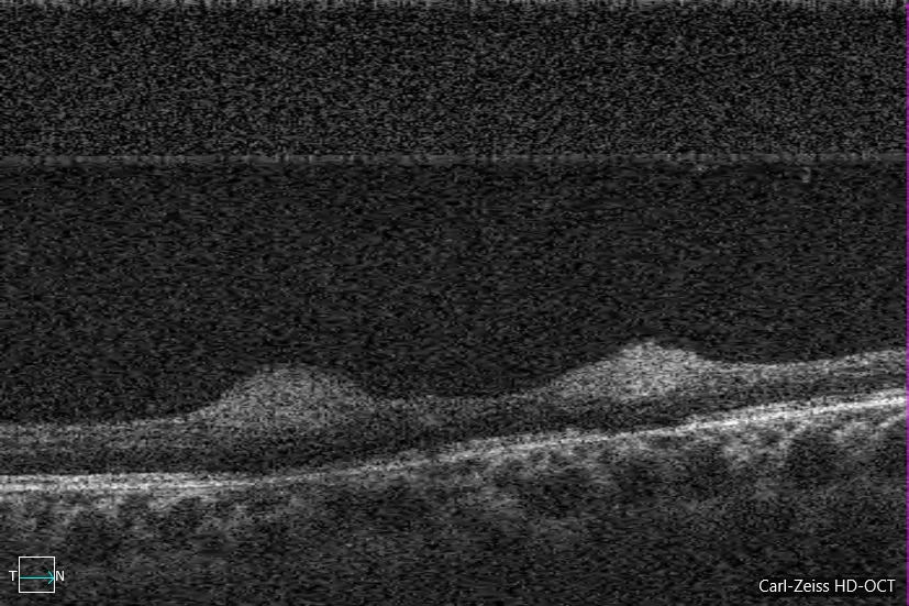
. Optical coherence tomography angiography (OCT-A) of the three retinal astrocytic hamartomas of the right eye. E and F: Horizontal B-scan over the temporal (E) and superonasal (F) hamartomas, showing segmentation for the superficial capillary plexus, where the lesions and their vasculature are located. G: OCT-A en face of the superficial plexus of the temporal retinal astrocytic hamartoma, showing a single feeding artery and a single draining vein, branching pattern.
. Optical coherence tomography angiography (OCT-A) of the three retinal astrocytic hamartomas of the right eye. E and F: Horizontal B-scan over the temporal (E) and superonasal (F) hamartomas, showing segmentation for the superficial capillary plexus, where the lesions and their vasculature are located. G: OCT-A en face of the superficial plexus of the temporal retinal astrocytic hamartoma, showing a single feeding artery and a single draining vein, branching pattern.
Description
Tuberous sclerosis is a rare genetic disease of autosomal dominant inheritance. It causes multiple hamartomas that appear in various organs such as the brain, heart, liver, kidneys and eyes. This tumor growth is caused by mutations in the TSC1 and TSC2 genes that code for hamartin and tuberin, proteins that decrease cell proliferation through the mTOR pathway. The diagnosis is clinical and is established through major criteria, such as retinal hamartomas, and minor criteria. mTOR inhibitors (Sirolimus and Everolimus) may decrease the appearance and growth of retinal hamartomas.