Tumors of the retina and choroid
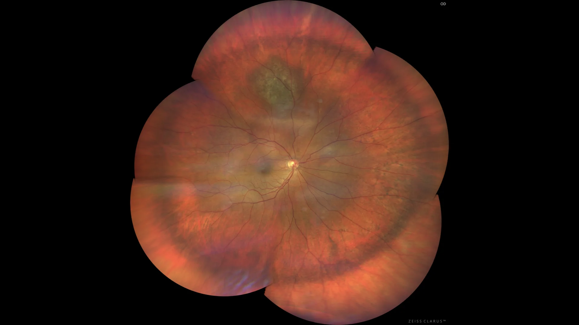
Color: raised pigmented lesion in the upper pre-equatorial region.
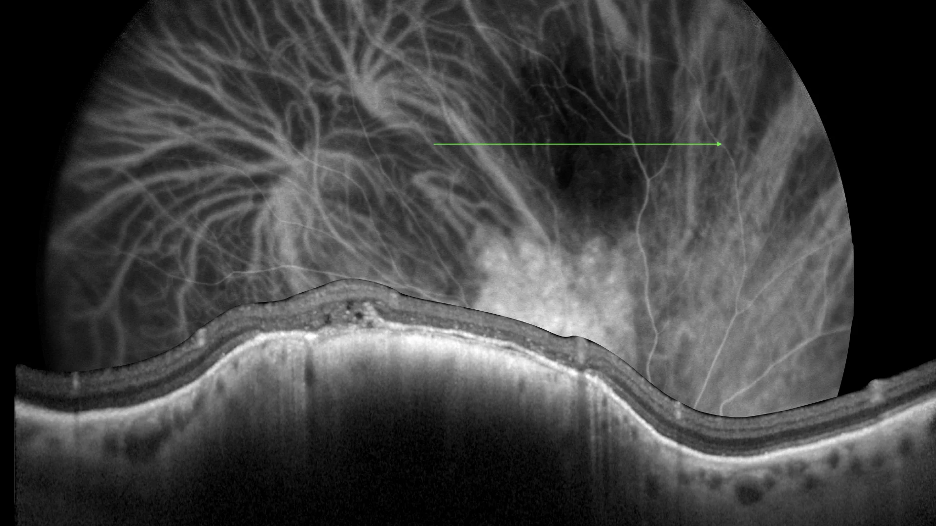
Indocyanine green angiography, showing a hypocyanescent lesion from early stages. SD OCT: raised lesion, with intense posterior block, characteristic of pigmented lesions of the choroid.
Description
Choroidal nevi are generally flat or slightly raised lesions, which may or may not be pigmented and are located in the choroid. They are usually no more than 2 mm thick and show a progressive increase from the choroid. They are often asymptomatic, although in rare cases they may cause flashes of light (in the presence of subretinal fluid) or a reduction in visual acuity (if located directly under the fovea). These characteristics are essential to distinguish a choroidal nevus from other ocular conditions, such as malignant melanoma of the choroid.