Tumors of the retina and choroid
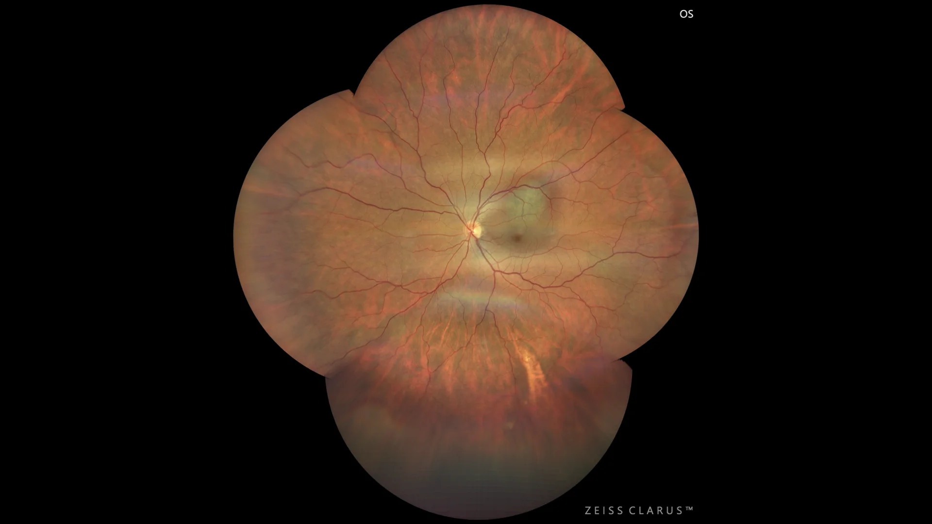
Wide-field color retinography with raised pigmented lesion measuring approximately 7 x 8 mm in diameter.
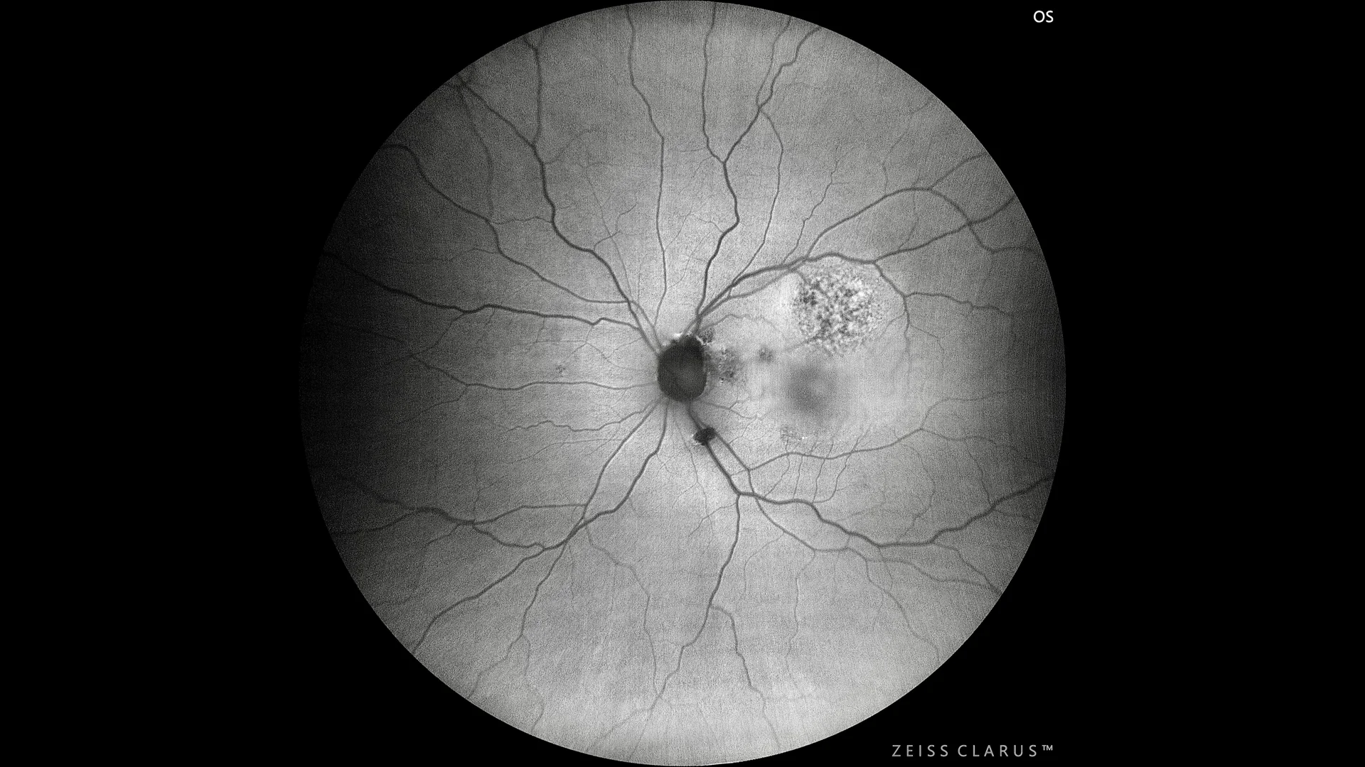
◦ Green autofluorescence: the lesion is shown with a mottled alteration of the pigmented epithelium
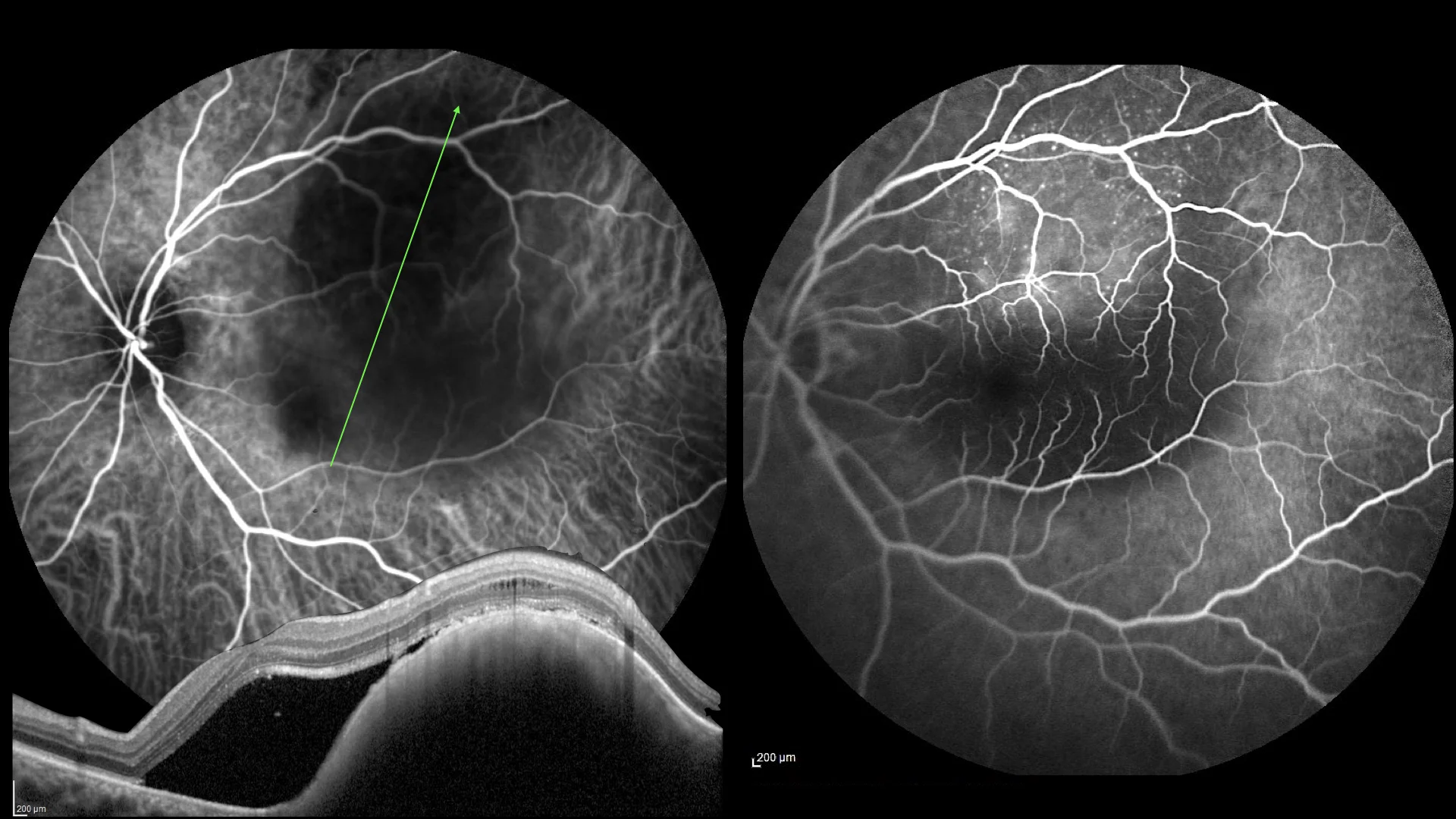
◦ OCT SD: raised lesion, with intense posterior block, abundant subretinal fluid, irregularity in the RPE, and fluid in the outer nuclear layer.
Description
Choroidal melanoma is a malignant neoplasm originating in the melanocytic cells of the choroid. This type of melanoma is the most common primary intraocular neoplasm in adults and may present with a variety of symptoms, including blurred vision, vision loss, flashes of light, and visual field changes. Diagnosis is based on a combination of ophthalmoscopic examination, ocular ultrasound, fluorescein-indocyanine green angiography, and optical coherence tomography (OCT). The classic appearance of a choroidal melanoma includes a raised tumor with variable pigmentation, often associated with serous retinal detachment. Treatment depends on the size and location of the tumor as well as vision in the affected eye and may include radiation therapy with brachytherapy plaques, external beam radiation therapy, surgical resection, or, in advanced cases, enucleation of the eye. Close follow-up is crucial to detect metastases, with the liver being the most common site of metastatic spread.