< back
Myopic retinoschisis
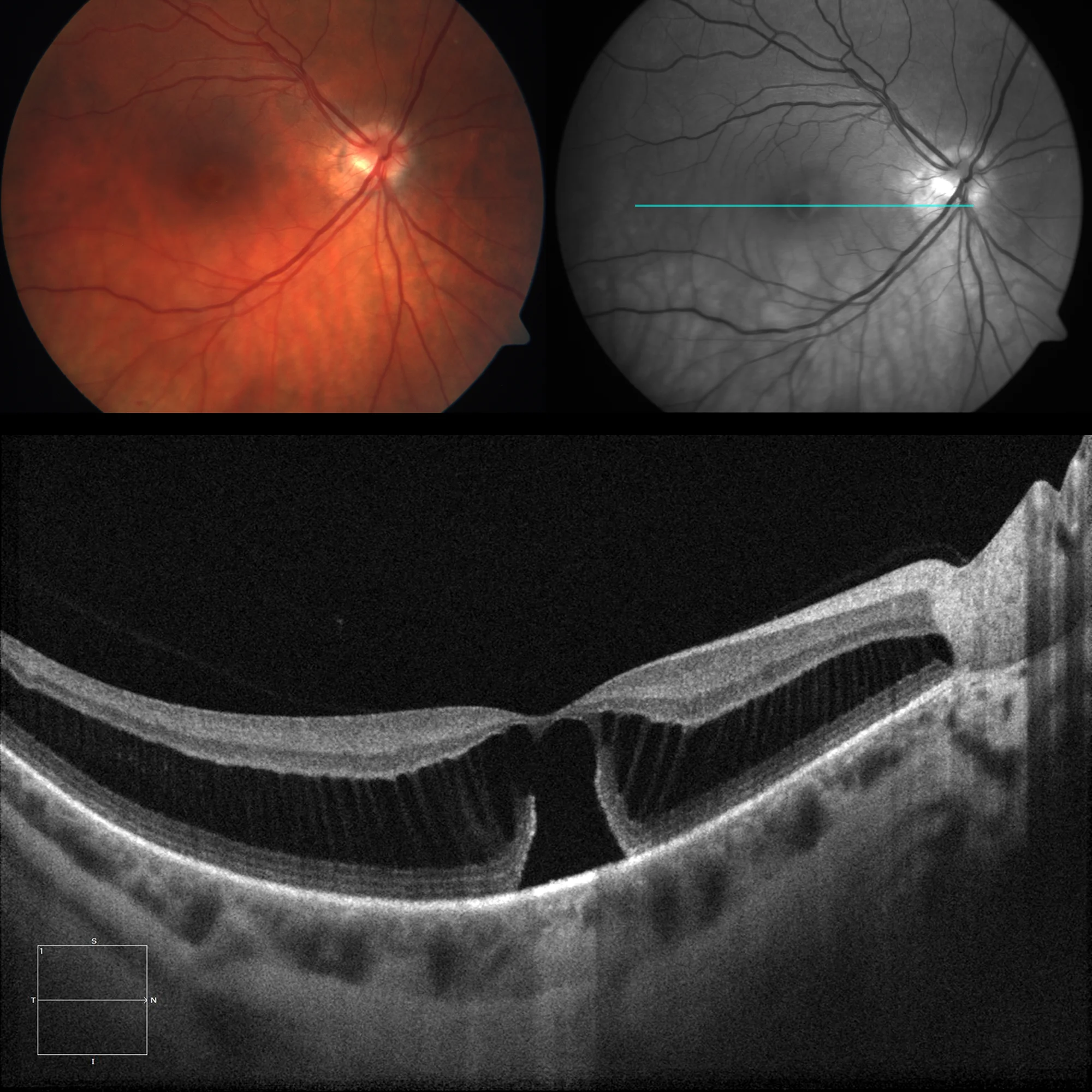
Color and green filter fundusography: Image of macular hole/pseudo-hole OCT 1 line HD: Marked macular retinoscopy with almost complete macular hole
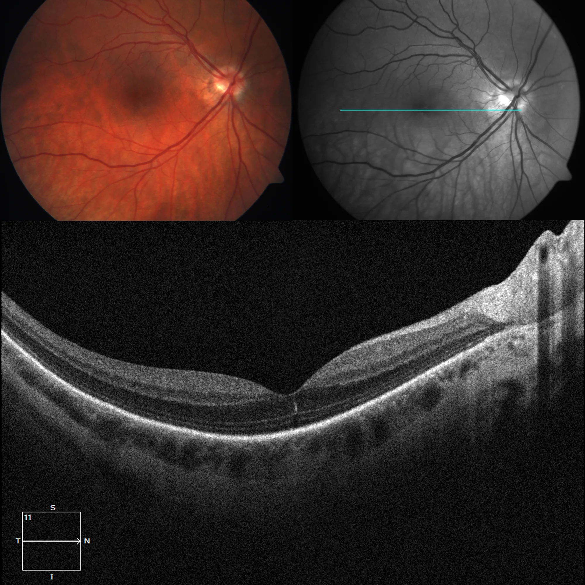
Color retinography and green filter: Normal-appearing macula OCT 1 line HD: Practically normal macula, with disappearance of retinoschisis.
Description
Myopic retinoschisis or foveoschisis was described by Takano in 1999. It is included within tractional myopic maculopathy. Myopic foveoschisis may present: epiretinal membranes, macular retinoschisis, lamellar or complete hole and foveolar detachment.