< back
Amelanotic choroidal nevus
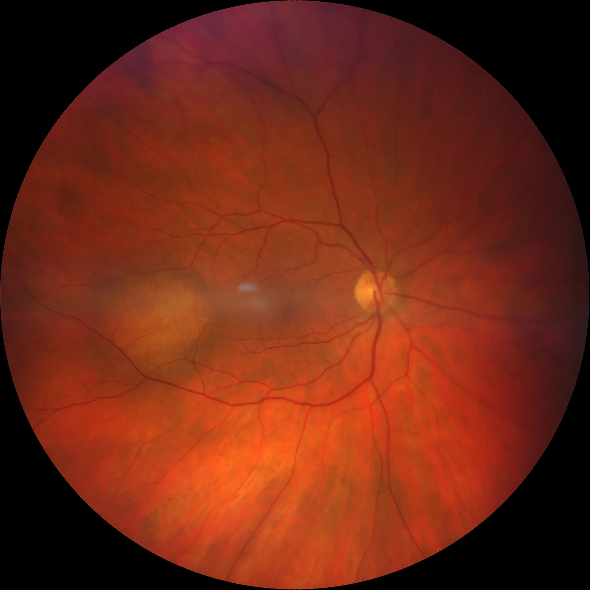
UWF Color Retinography: Temporary yellowish lesion OD.
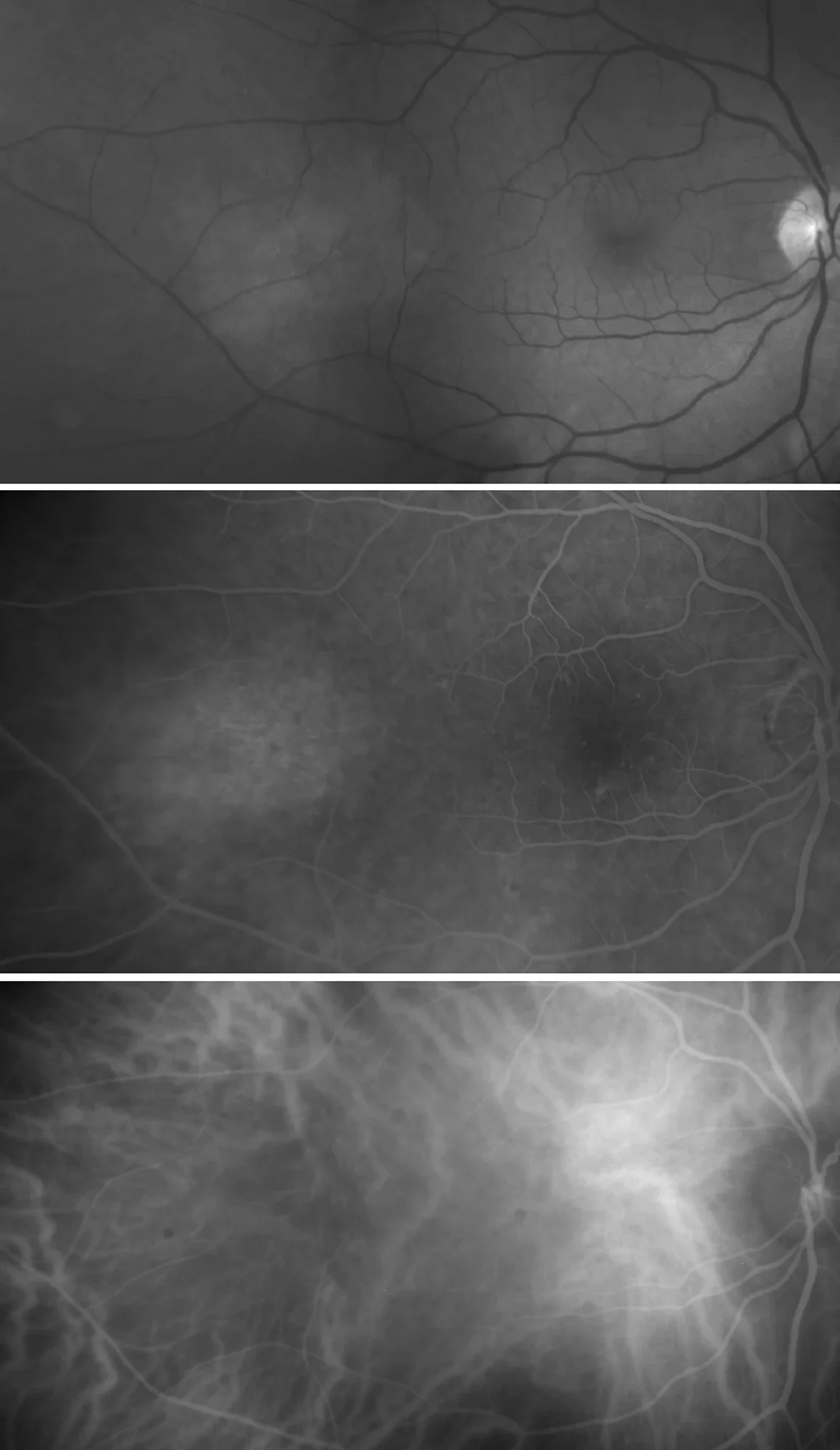
Green filter Clear lesion without vascular alterations AFG: Mild mottled hyperfluorescence in late stages ICGA Indocyanine green angiography without alterations
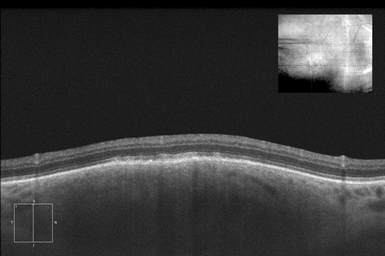
OCT HD: Almost flat choroidal lesion with mild RPE alteration and absence of fluid.
Description
Amelanotic choroidal nevi are rare benign tumors (5% of choroidal nevi), usually asymptomatic. The most important aspect is differentiation from other choroidal lesions.
Comments
Amelanotic nevus raises diagnostic doubts with choroidal lesions such as amelanotic melanoma, metastasis and choroidal hemangioma. Tests:- Widefield color retinography (Clarus 700, Zeiss): Yellowish rounded lesion in the temporal area of the right eye.
- Green filter (FF450 IR plus, Zeiss): Clear lesion without vascular alterations
- AFG (FF450 IR plus, Zeiss): Mild mottled hyperfluorescence at late times
- Indocyanine green angiography (FF450 IR plus, Zeiss): slightly hypocyanescent lesion without significant alterations
- Optical coherence tomography (Cirrus-HD 5000, Zeiss): Almost flat lesion with mild RPE alteration and absence of fluid.