Myopic neovascularization
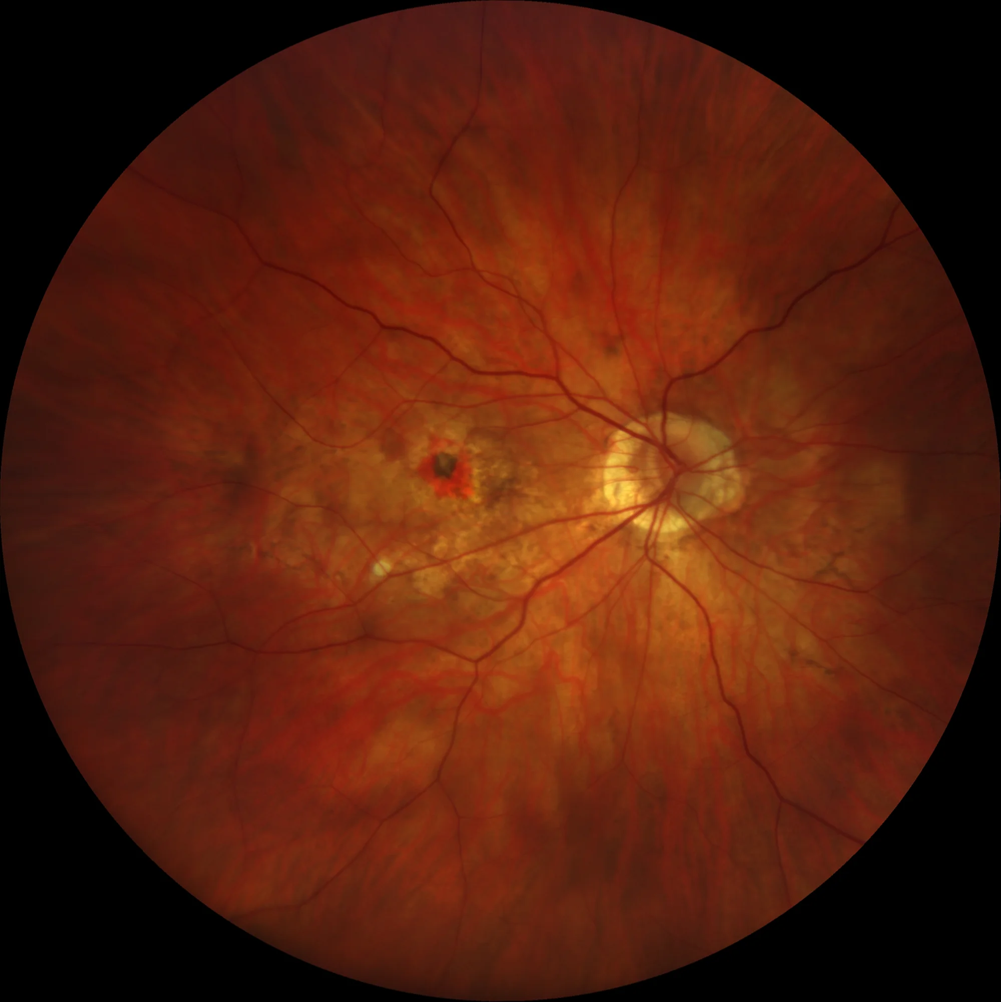
Pre-treatment retinal image OD: Pigmented juxtafoveal myopic neovascular membrane with perilesional hemorrhage. Peripapillary chorioretinal atrophy.
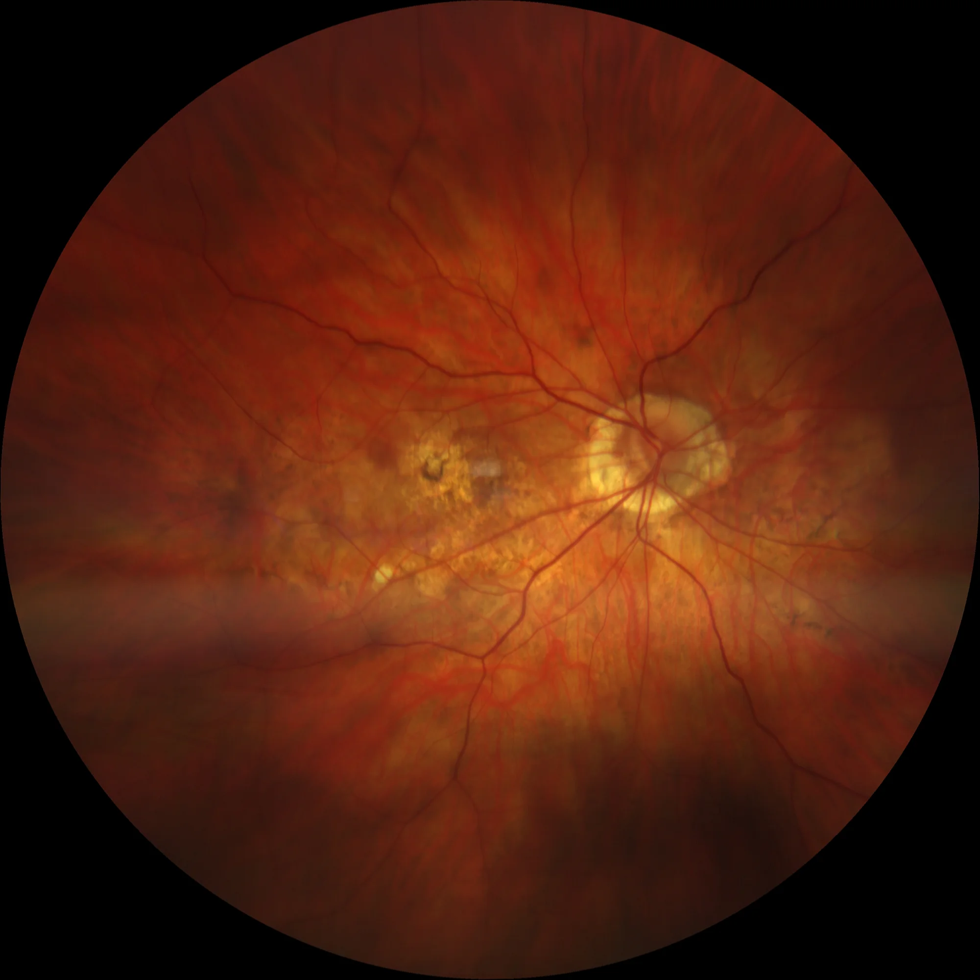
Post-treatment retinography OD: Juxtafoveal neovascular membrane with pigment at the edges and with perilesional pigmentary alteration.
Pre-treatment autofluorescence OD: In the autofluorescence we observe the hyperautofluorescent membrane surrounded by a hypoautofluorescent area that corresponds to the screen effect caused by the hemorrhage. Also notable in this image is the presence of a linear pigmentary alteration that runs below the macula from the optic nerve to the temporal area and that may be compatible with a lacquer streak.
Post-treatment autofluorescence OD: The scar lesion is observed as a small hypoautofluorescent zone surrounded by a hyperautofluorescent ring. The hypoautofluorescence related to the microhemorrhage has disappeared as the hemorrhage has been reabsorbed.
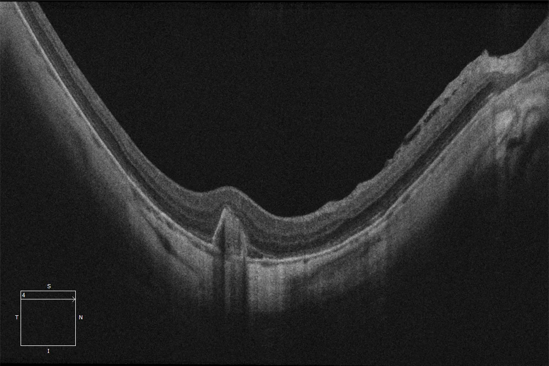
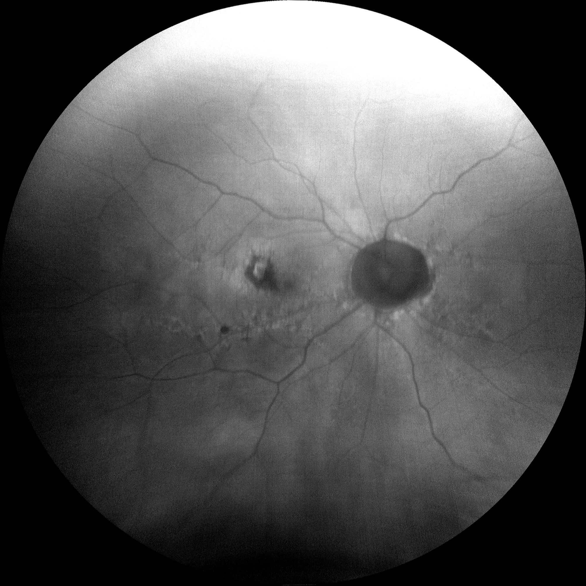
Macular OCT (slice over lesion) pre-treatment OD: We can see that the slice shown does not pass through the center of the fovea but through the center of the lesion which is slightly supero-temporal to it. The OCT shows the presence of a hyperreflective lesion above the RPE compatible with a type 2 neovascular membrane.

Macular OCT (foveal slice) pre-treatment OD: Maintains a preserved foveal profile with no signs of activity at the level of the fovea center.
Pre-treatment macular OCT-angiography OD (6 mm): OCT-A slice segmented at the level of the outer retina where the presence of a juxtafoveal subretinal neovascular complex appears. The hyperreflective lesion that appears in the image could reflect not only the neovascular complex but also the perilesional hemorrhage. In highly myopic patients, OCT-angiography is often subject to artifacts due to the difficulty in segmentation and the presence of other types of artifacts, which is why it is often difficult to visualize the neovascular membrane with this technique.
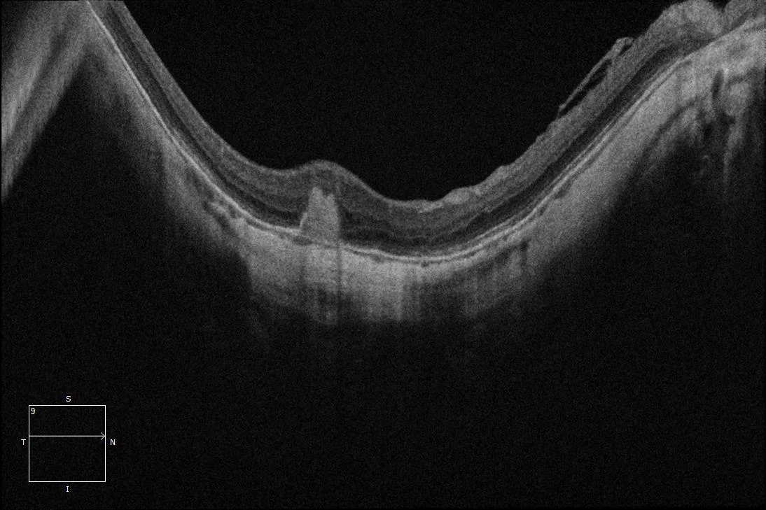
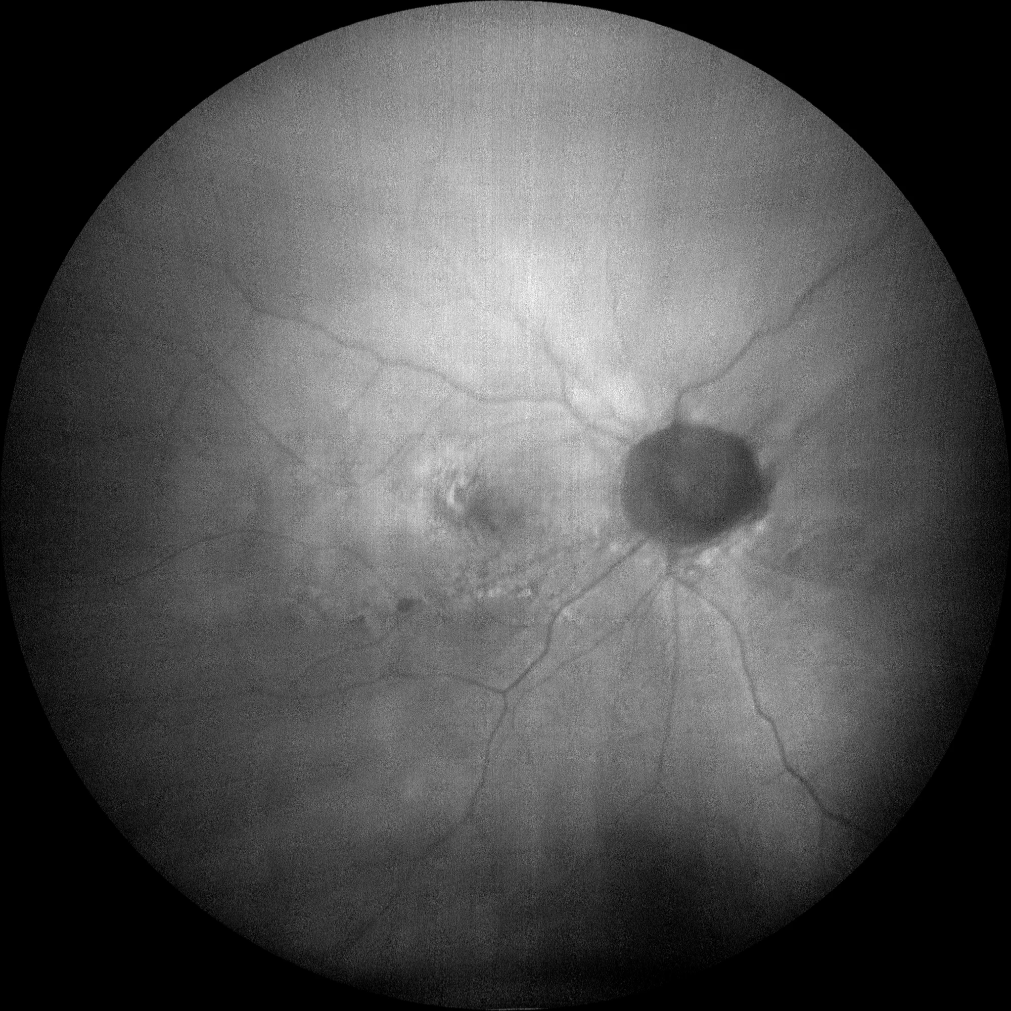
Post-treatment macular OCT OD: Subretinal hyperreflective lesion with no major changes compared to previous lesion.

Post-treatment macular angio-OCT OD (3 mm): In the section that segments the outer retina, a clear neovascular network is not observed but rather a zone of hyporeflectivity with some vessels at the edge of the lesion that does not correspond to neovessels, but rather to a projection artifact of the most superficial layers, since said vessels completely coincide in shape and size with the most superficial plexuses.
Description
It is defined as a neovascularization of choroidal origin (neovascular membrane) in the context of pathological myopia. Typically, they tend to be subretinal neovascular membranes (type 2 or classic) and are generally located in the foveal or juxtafoveal area, although they can also appear extrafoveal or peripapillary. In the fundus, they appear as a grayish lesion with pigmented edges, and may or may not be associated with retinal hemorrhage or exudation. Typically, these membranes can be active, giving rise to symptoms in the patient, without the presence of fluid, hemorrhage or exudation, so when deciding whether to treat the patient, the presence of symptoms is essential. In its evolution, three phases can be distinguished:
- Active phase: The membrane presents activity that is generally reflected in the appearance of symptoms by the patient and that may or may not be associated with the presence of retinal hemorrhage, fluid in the OCT or exudation.
- Scarring phase (Fuchs’ spot): Over time, a scarring process usually occurs, leading to pigmentation of the membrane and reduction of exudation.
- Atrophic phase: Occasionally, the membranes may develop chorioretinal atrophy around the neovascular lesion, leading to severe visual loss.