Aneurysmal type 1 neovascular membrane
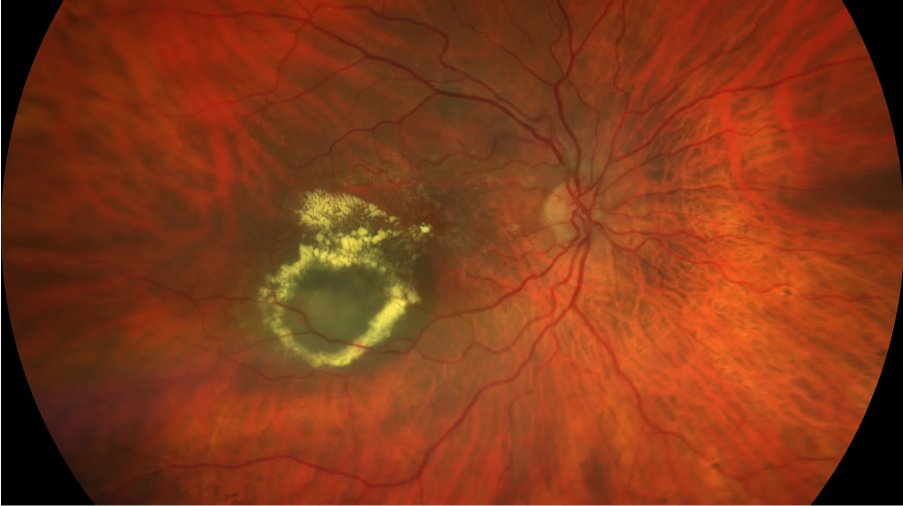
Color retinography (CLARUS 500, Zeiss): Marked lipid exudation temporal to the macular area in a circinate shape, coinciding with an underlying choroidal nevus. Presence of cuticular drusen.
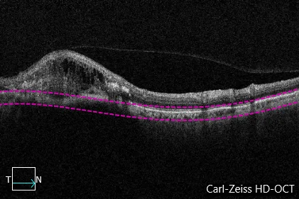
• OCT (CIRRUS 6000, Zeiss): Focal vitreomacular traction. At the temporal level there is cystic macular edema associated with small elevations of the pigmented epithelium, characteristic of the presence of aneurysmal dilatations. Choroidal shadow secondary to the presence of a nevus.
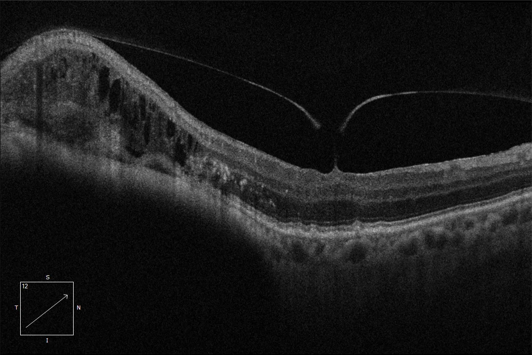
OCT: Focal vitreofoveal traction. Temporal cystoid macular edema overlying a detachment of the vascularized pigmented epithelium. Presence of a choroidal nevus.
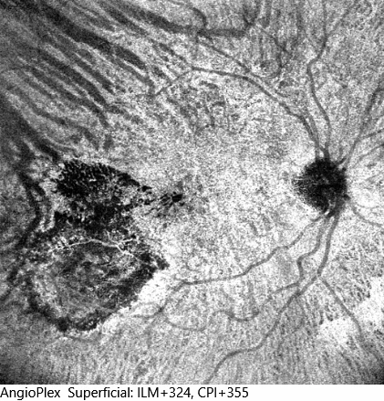
Angio-OCT (Cirrus 6000, Zeiss): The 12 x 12 mm image shows a neovascular complex with rounded structures in its bed compatible with aneurysmal dilatations.
Description
This is a subtype of exudative age-related macular degeneration characterized by a growth of the neovascular complex beneath the pigmented epithelium. Characteristically, they present aneurysmal dilatations (classically called polyps) in the vascular bed. This type of neovascular membranes usually generate greater exudation, reflected in the presence of multiple lipid exudates forming a circinated image. In other cases, the presence of subretinal hemorrhages is its main sign.