Torpedo maculopathy
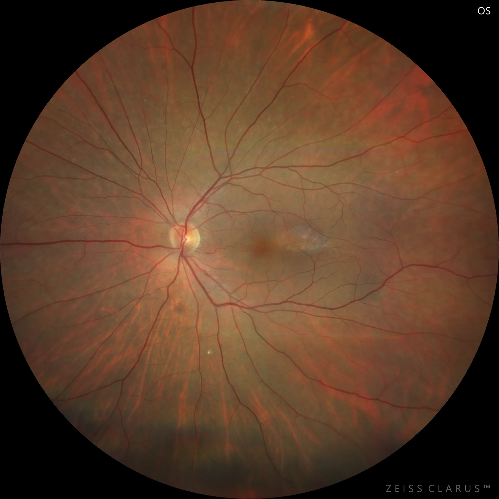
WF color retinography: Oval hypopigmented lesion temporal to fovea in LE.
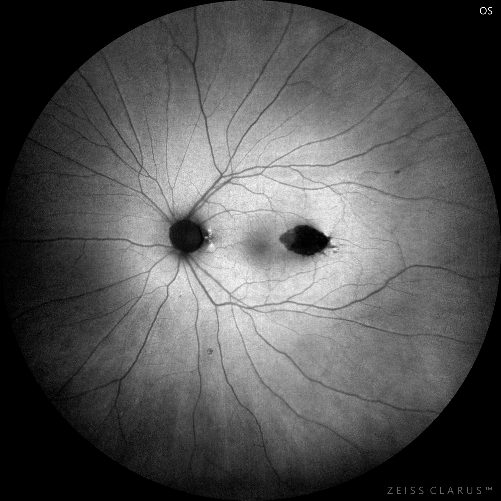
Background autofluorescence (AF Blue): Oval hypoautofluorescent lesion with well-defined edges and small hyperautofluorescent dots inside.
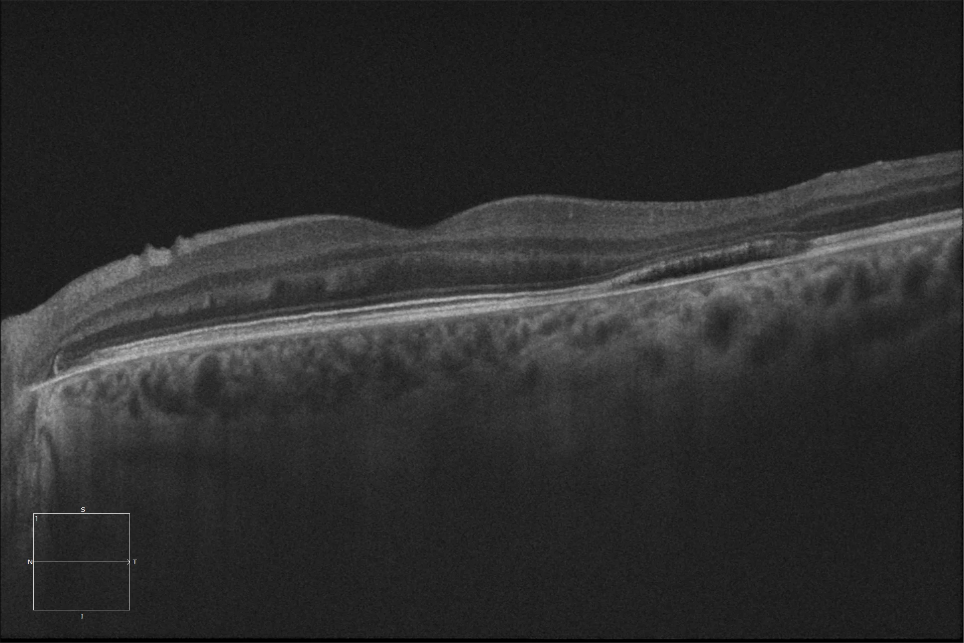
OCT HD: OCT shows cavitation in the outer layers of the retina, as well as loss of the ellipsoid zone (type 2 lesion), with increased transmission signal along the choroid.
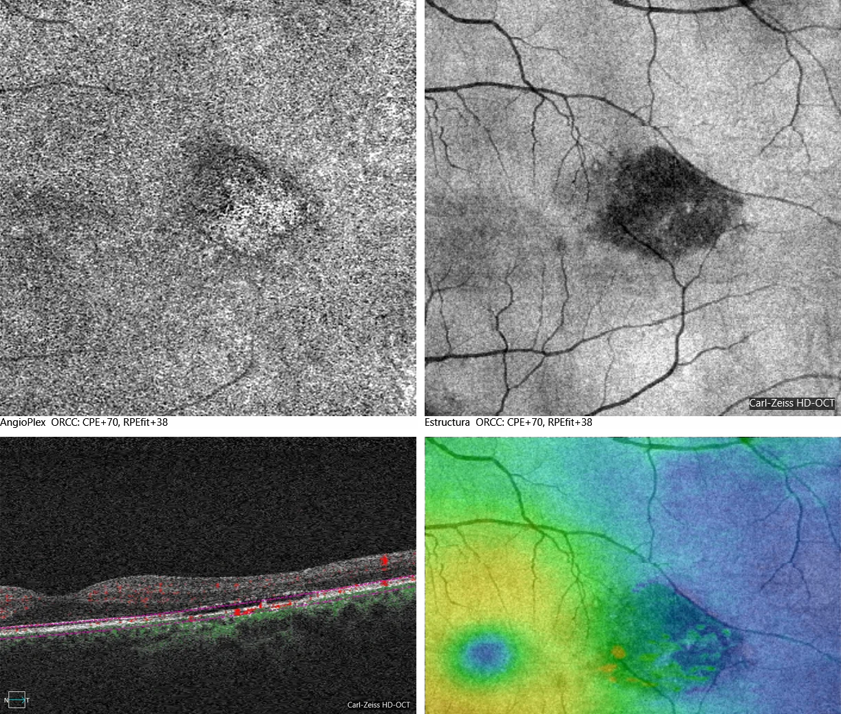
Anglioplex (Cirrus 5000): Oval lesion at the RPE level that allows visualization of choroidal vessels.
Description
Torpedo maculopathy is a congenital disorder that is usually unilateral and asymptomatic. The fundus image with an oval hypopigmented lesion temporal to the fovea, together with the phonograph imaging techniques (autofluorescence and OCT) provide the keys to the diagnosis.