Myopic macular hemorrhage
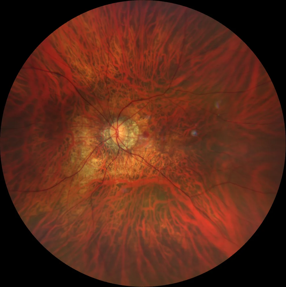
• Retinography (Clarus 700, Zeiss): central macular rounded hemorrhage. Tessellated fundus, peripapillary atrophy and diffuse chorioretinal atrophy affecting the macular area.
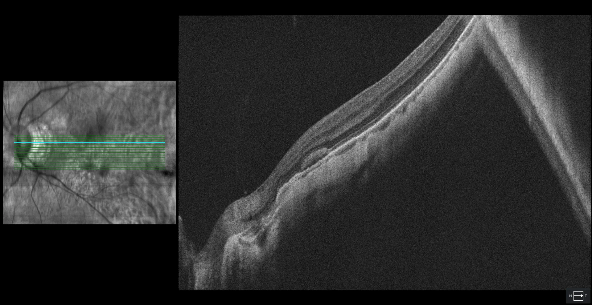
MHSR with a hyporeflective space separating it from the EPR
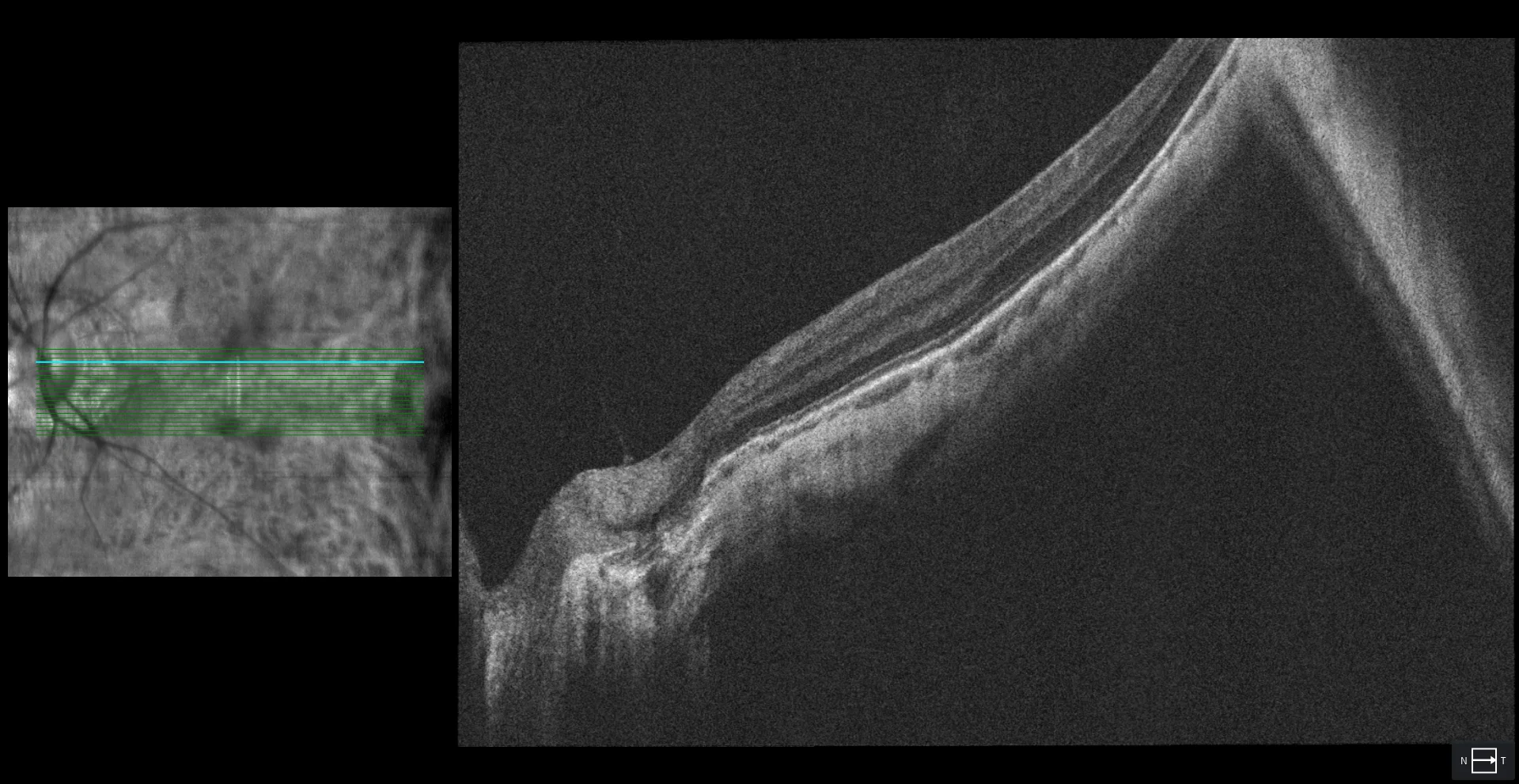
This suggests that this is a spontaneous macular hemorrhage and not a VAP. Angio-OCT also rules out the presence of a VAP (not shown). After 2 months of follow-up, the VAP has been spontaneously resorbed and vision has improved.
Description
A 47-year-old male comes to the doctor due to sudden loss of vision in his right eye.
AV OD 20/40 with -9.00 -4.25 x 160º.
Fundus examination reveals a central macular rounded hemorrhage. Tessellated fundus, peripapillary atrophy, and diffuse chorioretinal atrophy involving the macular area are also seen. OCT reveals subretinal hyperreflective material (SRHM). After 2 months of follow-up, the hemorrhage was spontaneously reabsorbed and vision improved to 20/20.