Vanishing hemorrhage
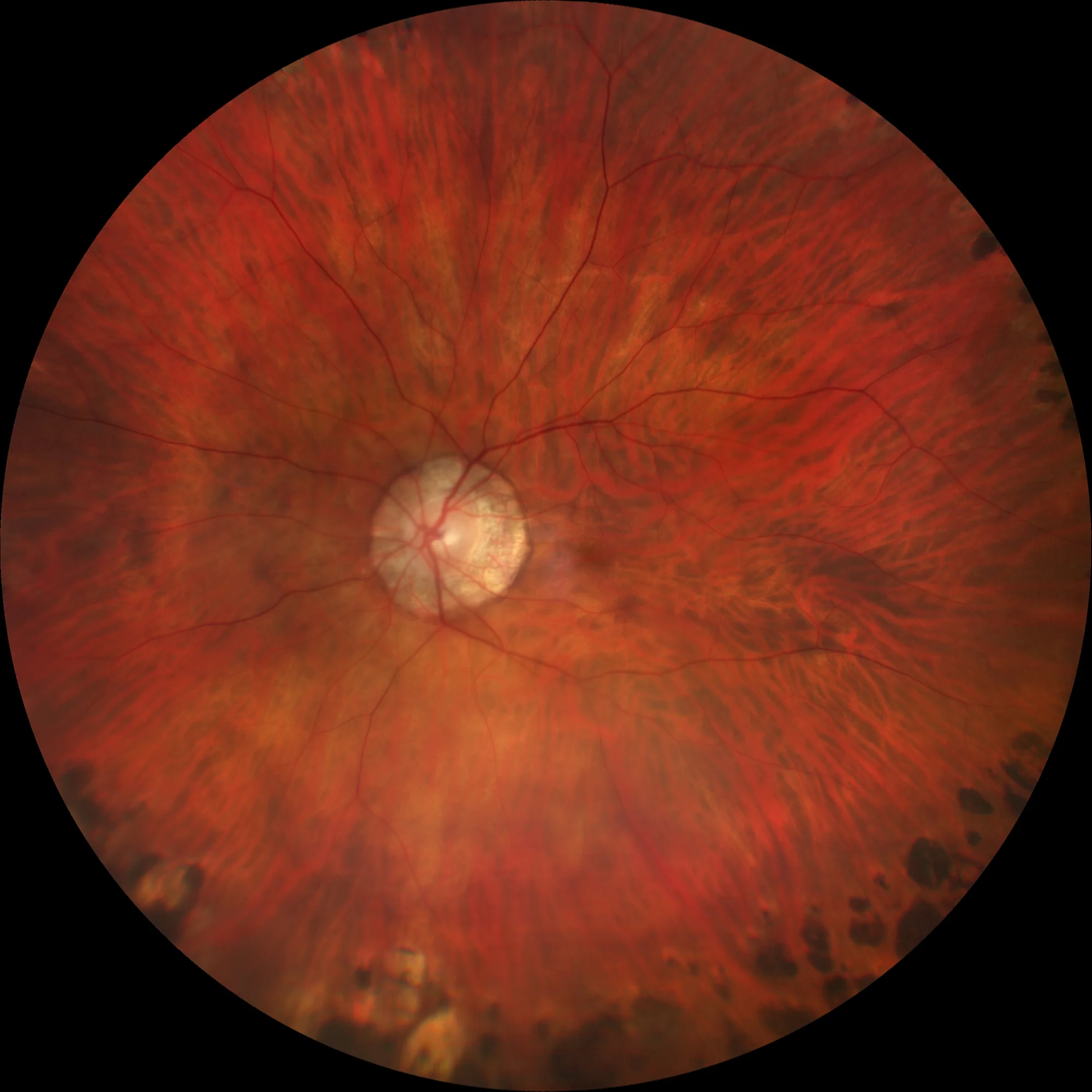
Retinography OD: High myopic fundus with lacquer streak in macular area. A small evanescent hemorrhage is observed infero-temporal to the macula. Laser impacts are also seen in the middle temporal periphery.
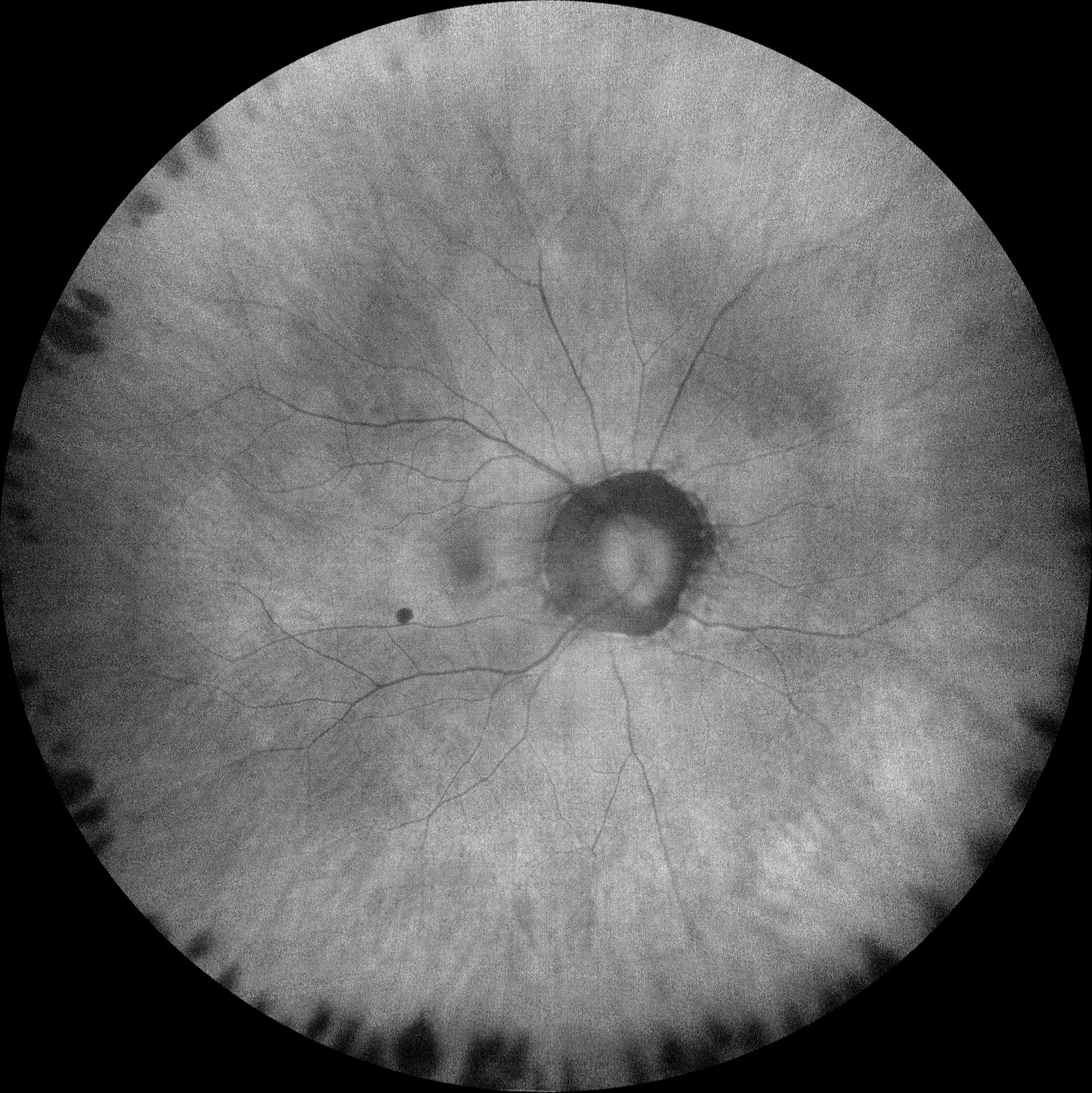
Autofluorescence OD: The hemorrhage stands out as a hypoaufluorescent (black) lesion due to the screening effect it produces.
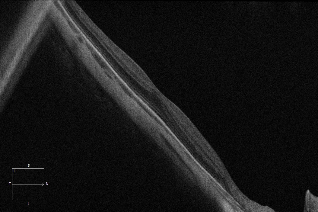
Macular OCT OD: Inclined macular plane due to macular staphyloma with preserved foveal profile.
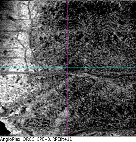
OCT angiography over the lesion: No neovascular membrane is observed on OCT angiography.
Description
Evanescent hemorrhages in myopes are usually subretinal hemorrhages (sometimes intraretinal) that occur during the development or expansion of a lacquer streak or rupture of the coricapillary complex-Bruch’s membrane. These hemorrhages may be located in the macula or extramacular. If they are located in the macula, they may be associated with vision loss that is generally transient and recovers when the hemorrhage disappears, so they do not require treatment. It is important to differentiate them from hemorrhages secondary to neovascular membranes that do require treatment with antiangiogenics.