Bergmeister papilla
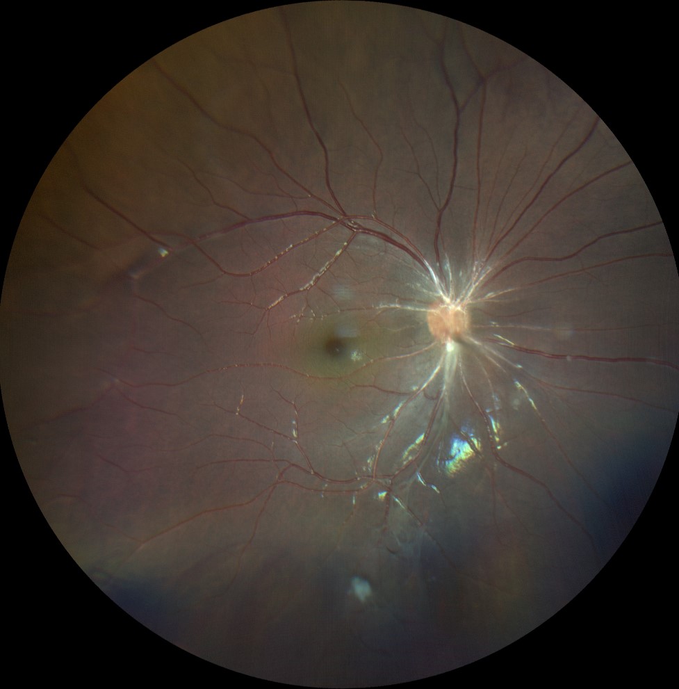
Color retinography (Clarus 500, Carl Zeiss) of the right eye, showing a fibroglial tract arising from the papilla and continuing anteronasal in the direction of Cloquet's duct.
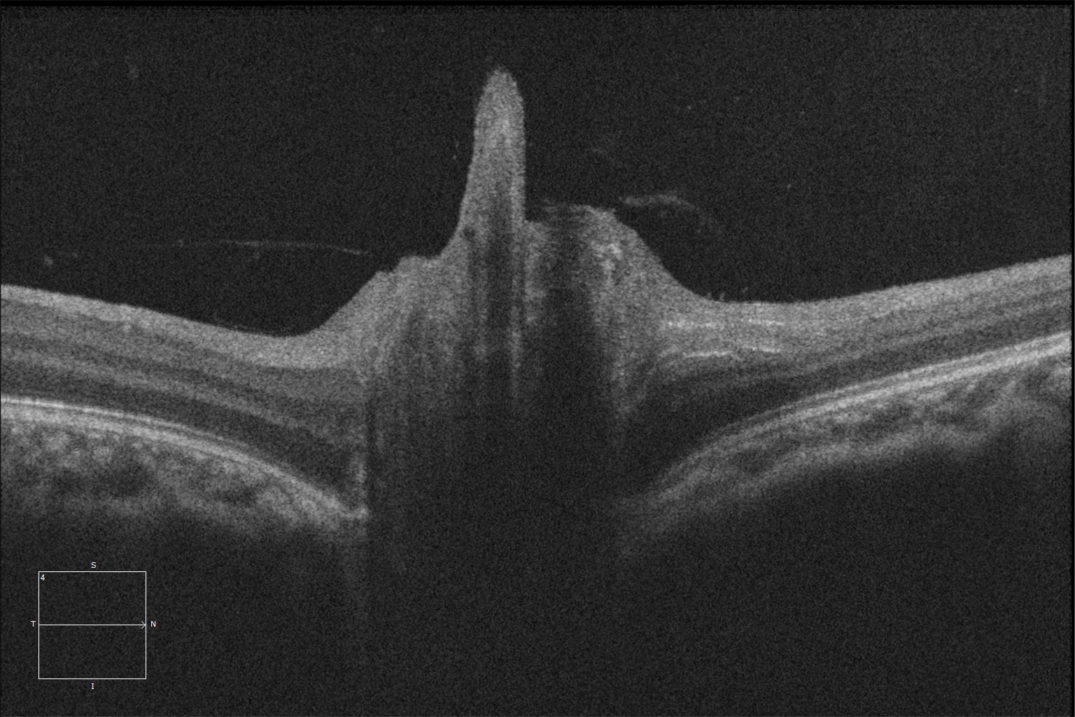
Structural optical coherence tomography at the level of the right optic disc (Cirrus 5000, Carl Zeiss Meditec ASG, Jena, Germany) showing this intermediate intensity tract emerging from the center of the optic disc of the right eye, in an anterior direction.
Description
The papilla of Bergmeister is a remnant of the fibrous sheath that covers the fetal hyaloid artery during embryonic development. Normally, this vessel atrophies after 30 weeks of gestation, giving rise to the hyaloid canal or Cloquet’s canal.
This is a mild variant of persistent fetal vasculature, which is usually asymptomatic and diagnosed incidentally.
The diagnosis is based on the visualization of glial and fibrous tissue extending anteriorly from the optic disc. Diagnostic tests such as optical coherence tomography (OCT) with angiography can be used to support the diagnosis, revealing a tract of intermediate reflectivity with possible flow signal emerging from the optic disc.
Treatment is not indicated in the absence of complications. Some patients may develop vitreoretinal traction or macular retinoschisis.