Compendium of clinical cases through multimodal imaging for the management and evolution of retinal ocular pathologies.
Cases
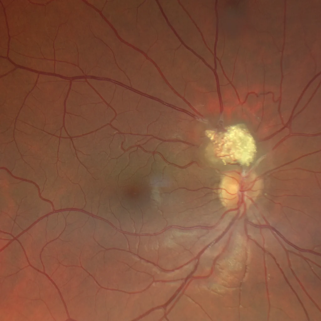
Retinal astrocytic hamartoma
Retinal astrocytic hamartoma
Retinal astrocytic hamartoma is a glial, vascularized, and benign tumor, usually asymptomatic. It typically presents as
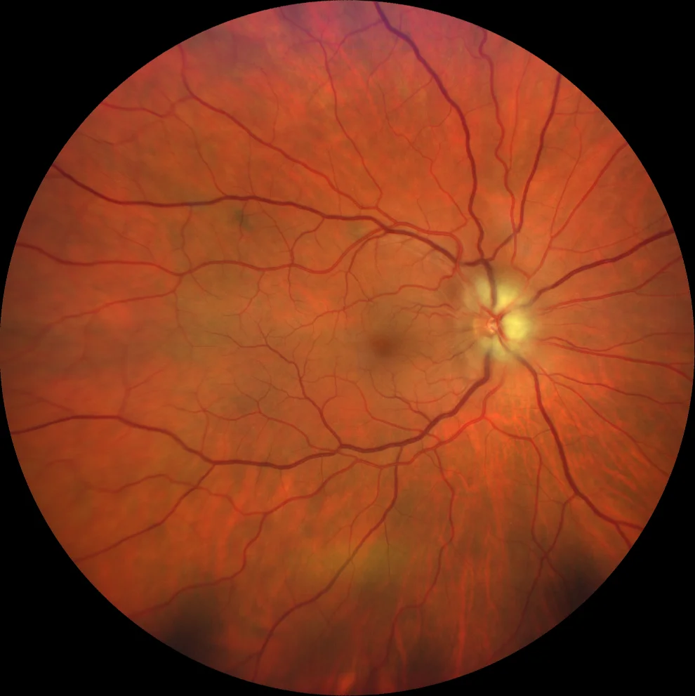
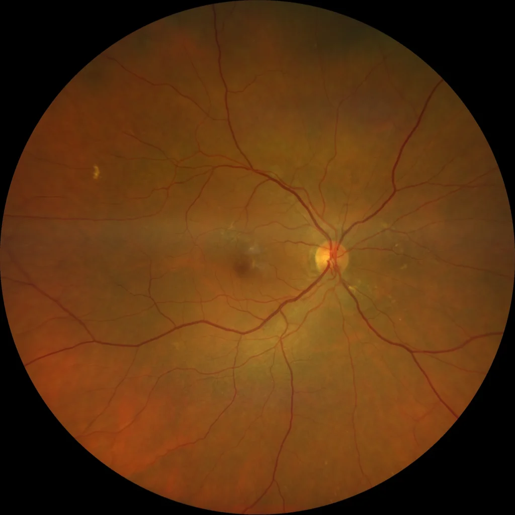
Anterior ischemic optic neuropathy – Non-arteritic
A 53-year-old woman, hyperopic (spherical equivalent OD +7.00 OI +6.50), complained of pain and red eye in the right eye that had been developing for
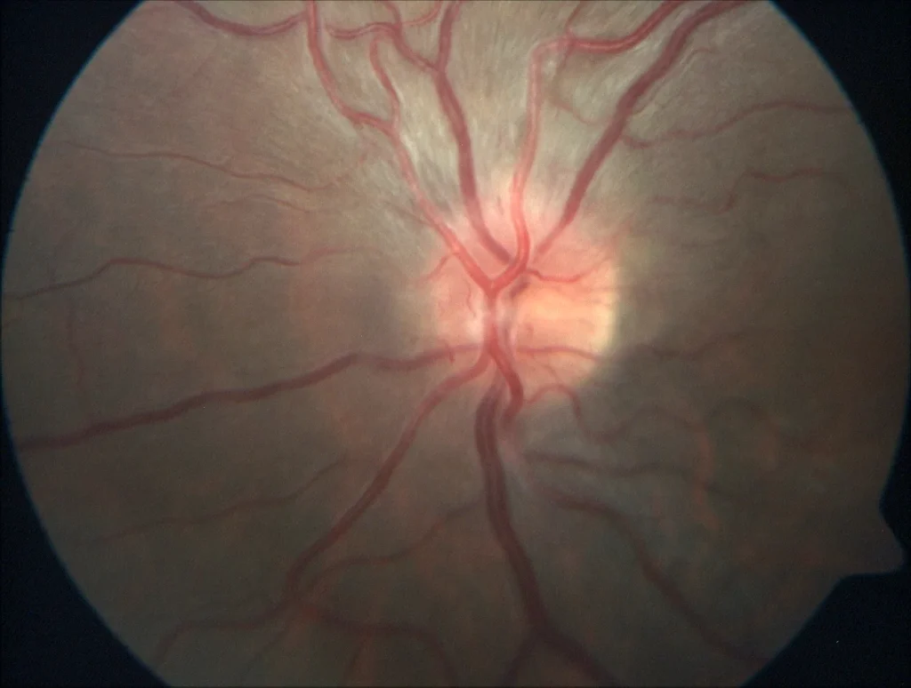
Unilateral anterior neuritis
Unilateral anterior neuritis
Optic neuritis is most common between the ages of 18 and 45, and usually presents with unilateral vision loss, color vis
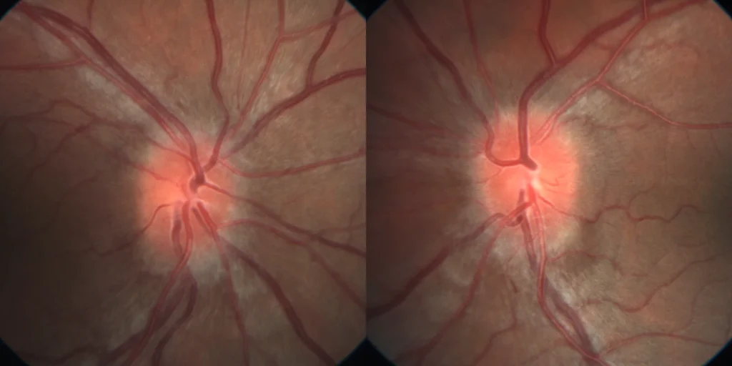
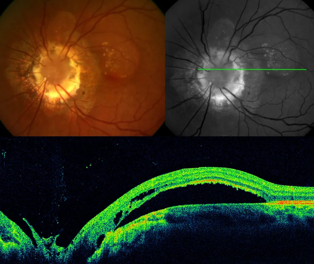
Papilledema due to idiopathic intracranial hypertension
Papilledema due to idiopathic intracranial hypertension
The presence of bilateral papilledema due to increased intracranial pressure is called papill
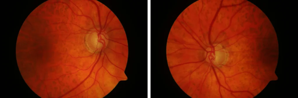
Advanced chronic simple glaucoma
Chronic simple glaucoma (CSG) is a progressive optic neuropathy secondary to increased intraocular pressure that causes visual field loss that can lea
Macular serous detachment associated with “morning glory”
Morning glory syndrome is an abnormality of papillary development that is frequently associated with intracranial abnormalities. 38% of cases present
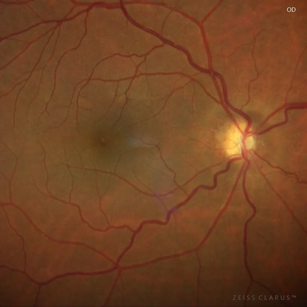
Optic nerve hypoplasia
Optic nerve hypoplasia is characterized by a small optic disc with a reduced number of axons. It can be unilateral or bilateral, and is frequently ass
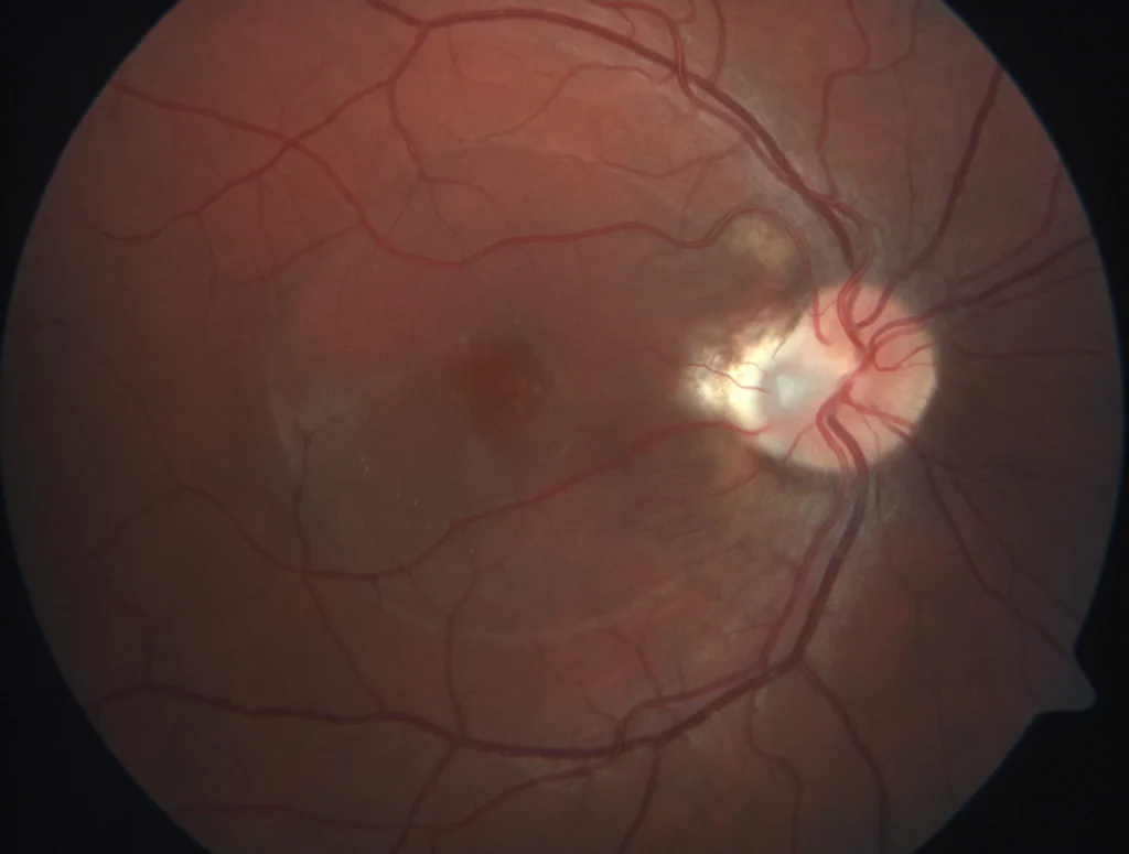
Optic pit with macular serous detachment
Papillary pit is a rare, usually unilateral, congenital abnormality of the optic disc that presents as a rounded, grayish temporal depression. Visual
Pachychoroid Neovasculopathy
Pachychoroid neovasculopathy. The spectrum of pachychoroid diseases refers to a group of conditions that share the pachychoroid phenotype, represente