Adult-Onset Foveomacular Vitelliform Dystrophy
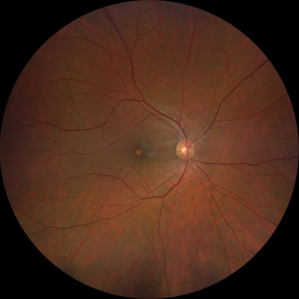
Retinography (Clarus 700, Zeiss): small yellowish deposit at central level (A1, A2).
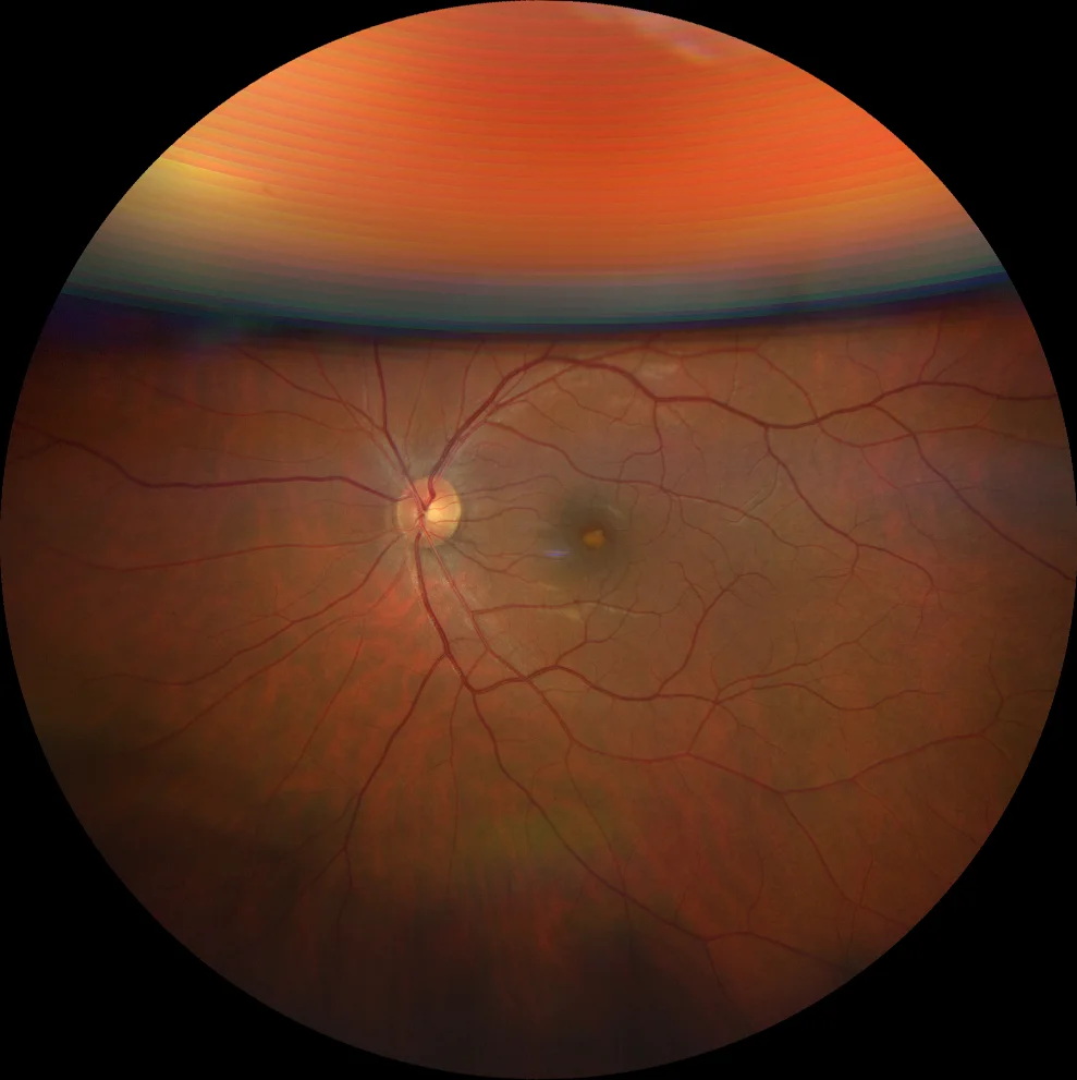
Retinography (Clarus 700, Zeiss): small yellowish deposit at central level (A1, A2).
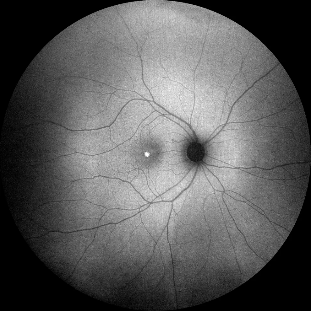
Green autofluorescence (Clarus 700, Zeiss): the deposits are hyperautofluorescent, indicating that they are vitelliform material (lipofuscin) (B1, B2).
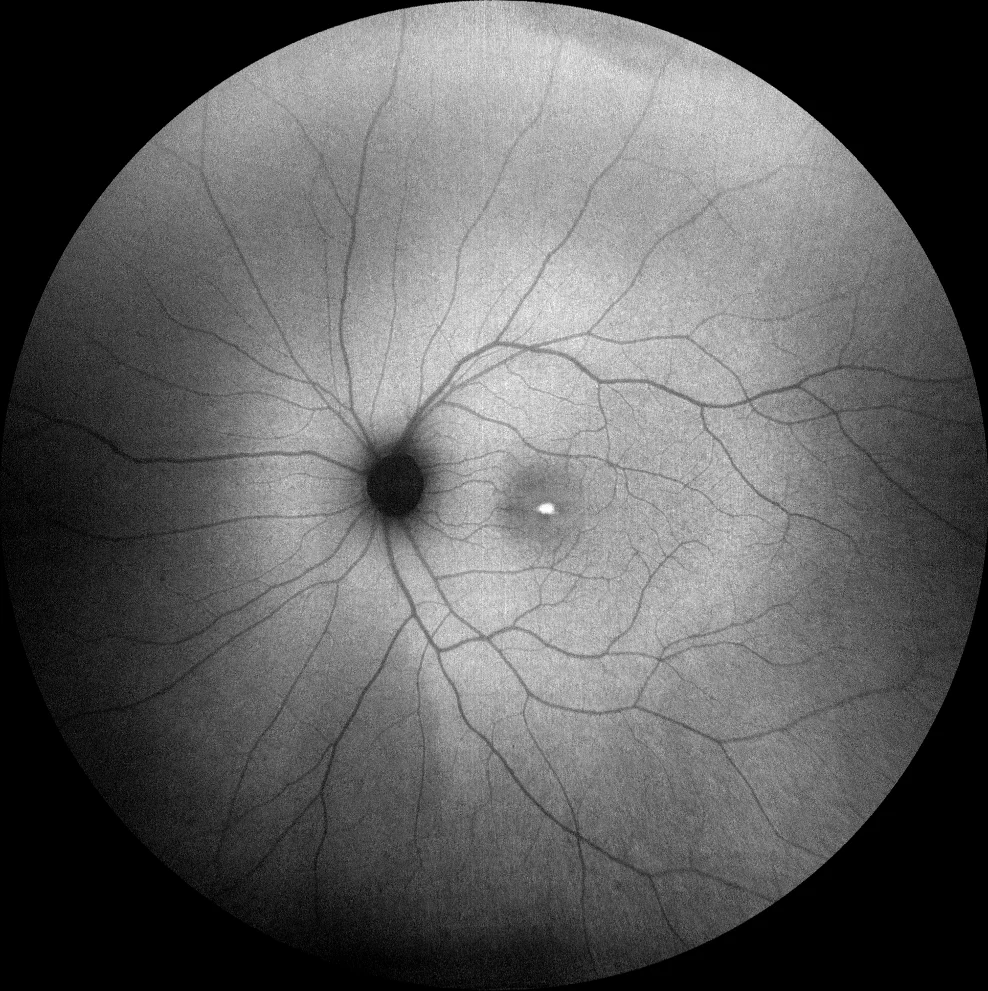
Green autofluorescence (Clarus 700, Zeiss): the deposits are hyperautofluorescent, indicating that they are vitelliform material (lipofuscin) (B1, B2).
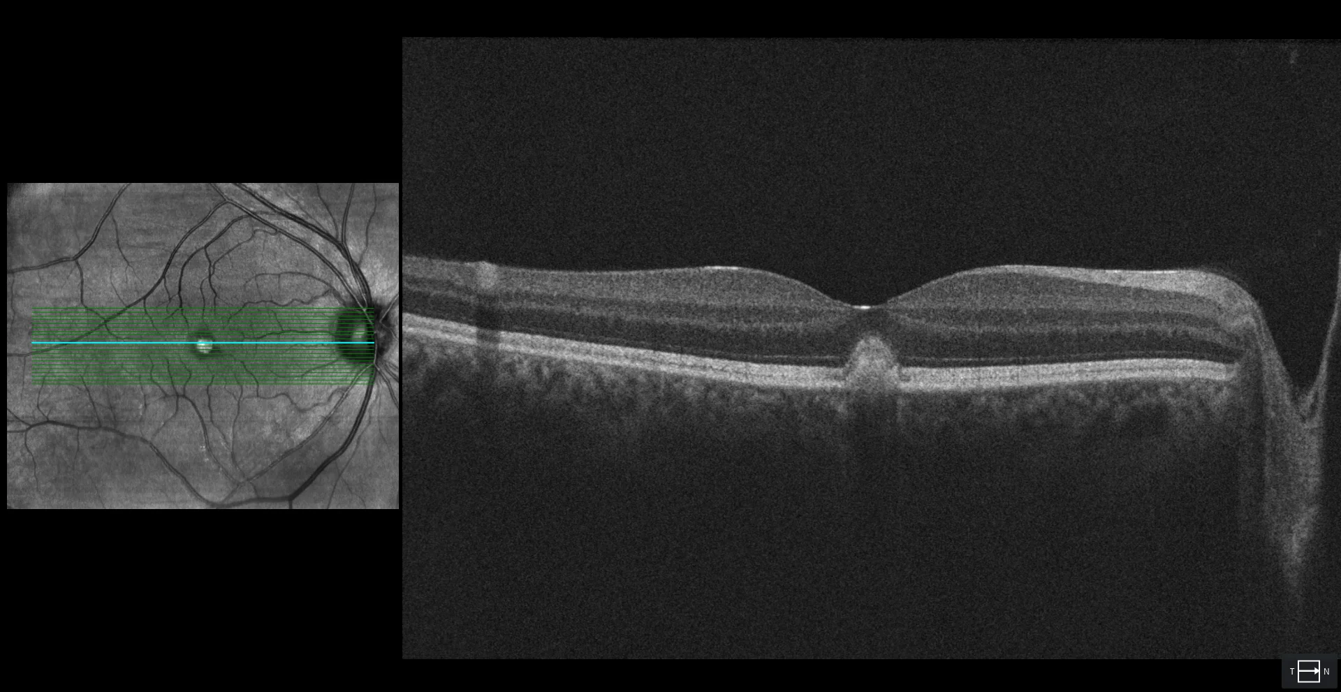
OCT (Cirrus 5000-HD, Zeiss): subfoveal hyperreflective material with preservation of the outer layers of the retina (EZ and ELM) (C1, C2).
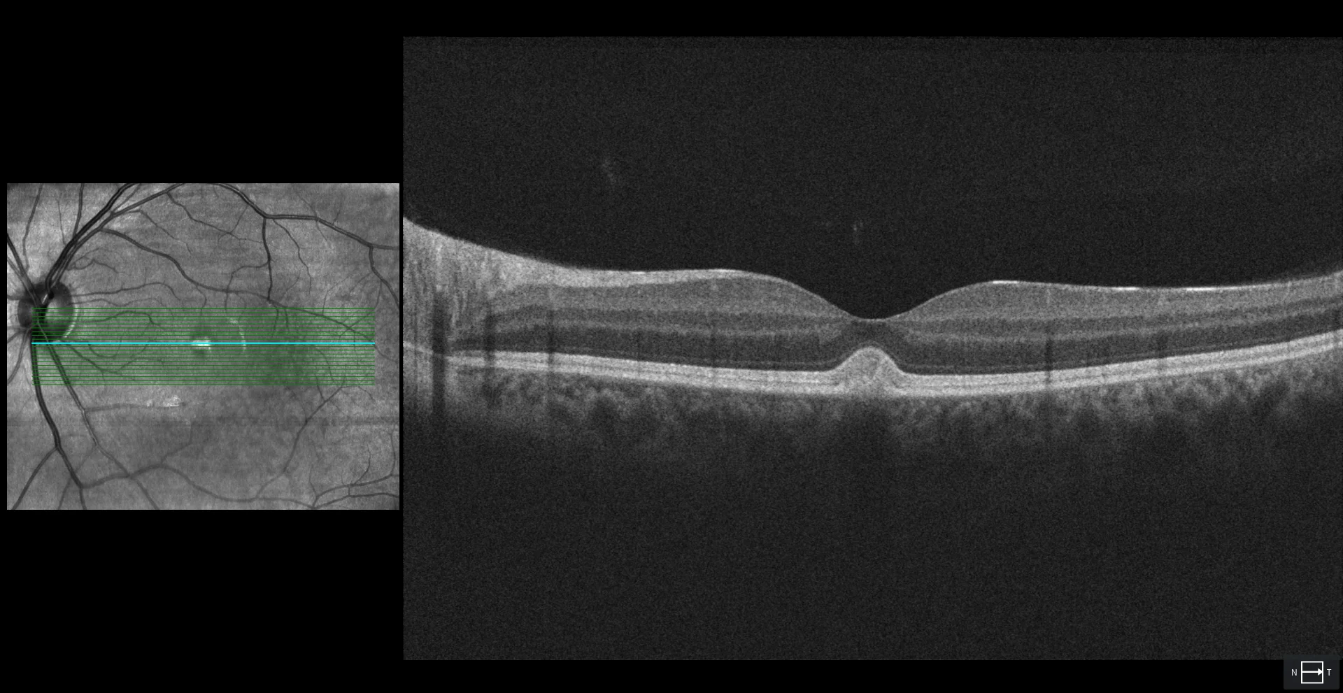
OCT (Cirrus 5000-HD, Zeiss): subfoveal hyperreflective material with preservation of the outer layers of the retina (EZ and ELM) (C1, C2).
Description
35-year-old male who comes in for a checkup.
VA OD 20/20 OS 20/20.
Fundus examination reveals a rounded yellowish lesion at the foveal level in both eyes, approximately 1/4 disc diameter in size. This deposit is hyperautofluorescent, suggesting the presence of vitelliform material. OCT shows hyperreflective material at the subfoveal level. EOG is normal, leading to a diagnosis of adult-onset foveomacular vitelliform dystrophy (AOFVD).