AMD
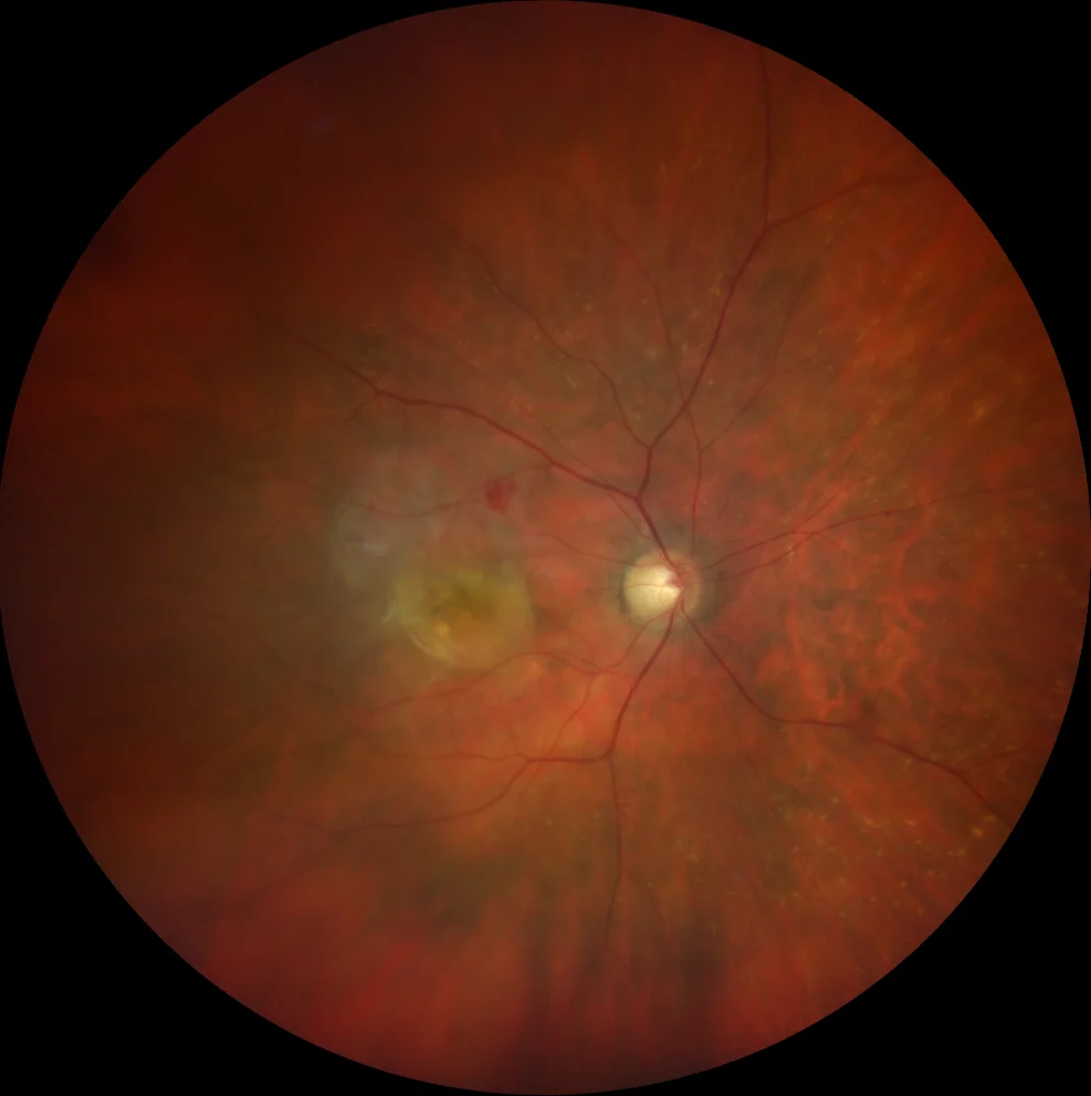
large yellowish macular lesion with a small hemorrhage above indicating activity.
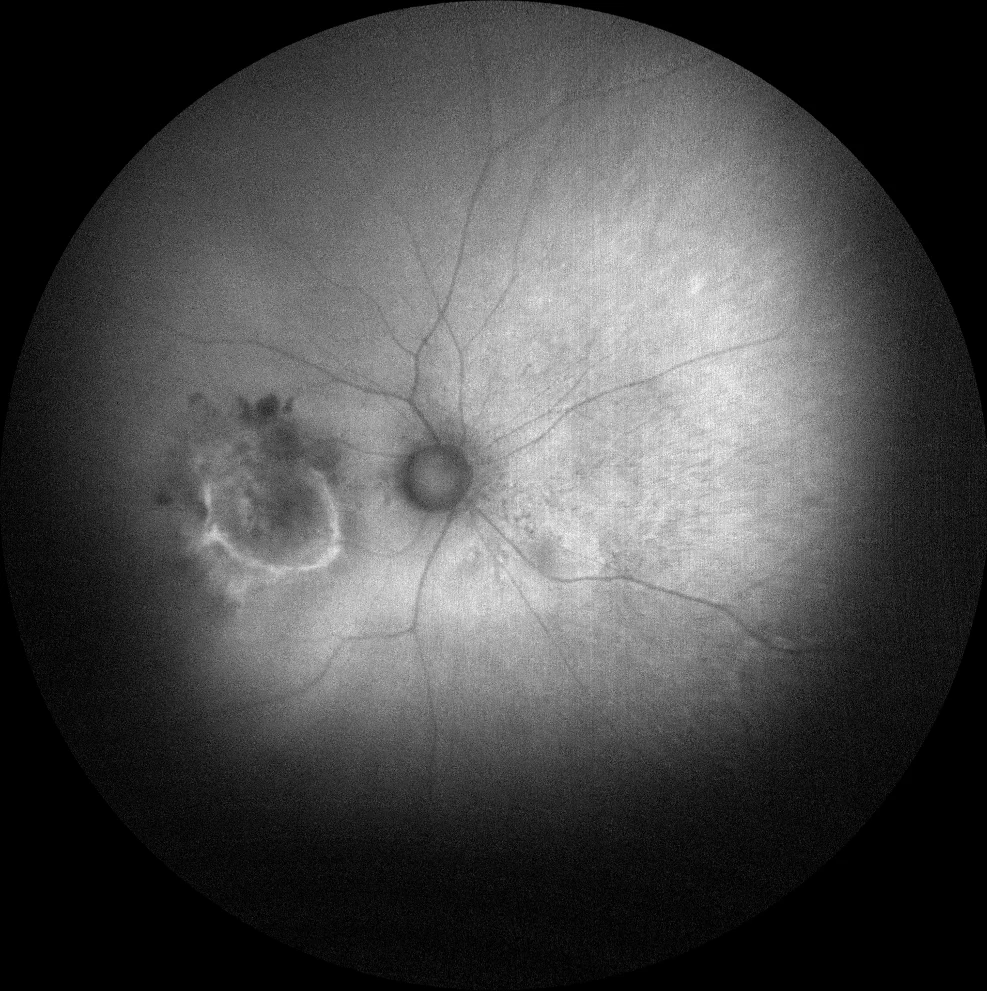
o B: The hyperautofluorescent edges of the lesion probably indicate an RPE with metabolic stress that will evolve towards atrophy and/or new areas of fibrosis.
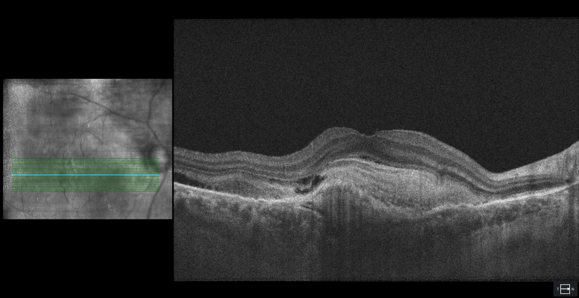
o C: subretinal hyperreflective material with a multilaminar structure corresponding to the different layers of fibrotic tissue that have formed in the subretinal space. A minimal amount of subretinal fluid is also observed at the temporal level, which also indicates lesion activity.
Description
78-year-old woman comes for a check-up.
The VA in RE is counting fingers and in LE 20/25.
The fundus in the RE shows a yellowish macular lesion and a small upper hemorrhage. OCT confirms the diagnosis of disciform scar secondary to neovascular AMD. The LE is being monitored for non-exudative type 1 NVM.