AMD Case 1 RPE tear
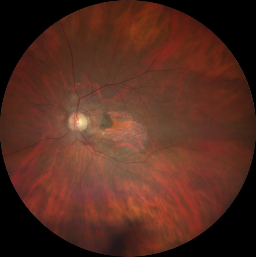
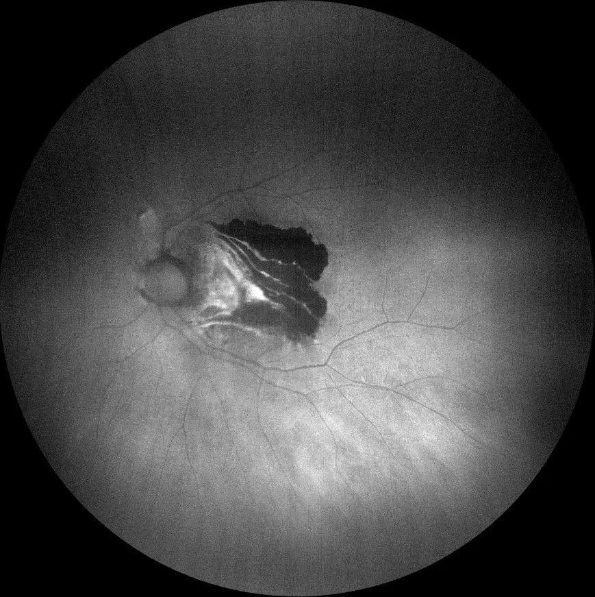
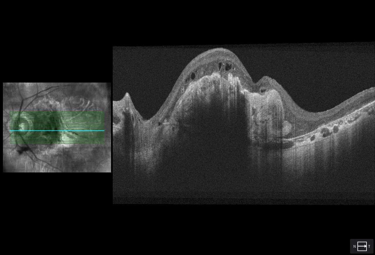
B1: RPE rupture. Choroidal vascularization can be seen in the area where there is no RPE and a dark area towards the nasal that corresponds to the coiled RPE.
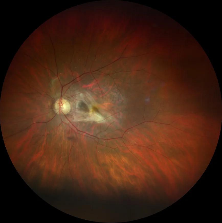
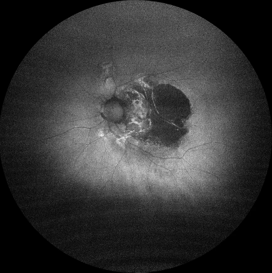
C1: onset of fibrosis after 6 months of follow-up
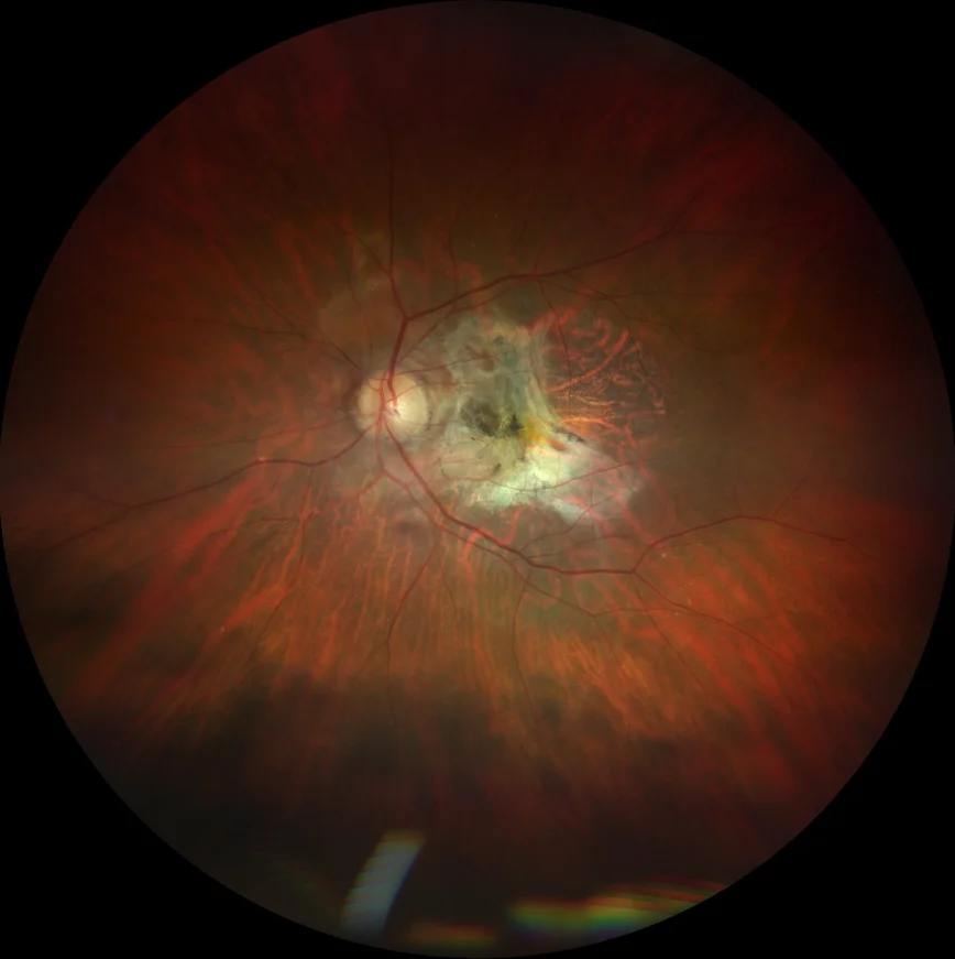
D1: severe macular fibrosis with pigment deposition after 2 years of follow-up
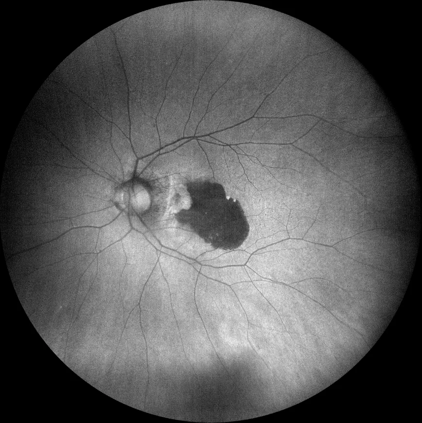
B2 (blue AF): The area where there is no RPE is hypoAF while the coiled RPE is seen as a hyperAF zone.
C2 (green AF): appearance after 6 months of follow-up
D2 (green AF): appearance after 2 years of follow-up
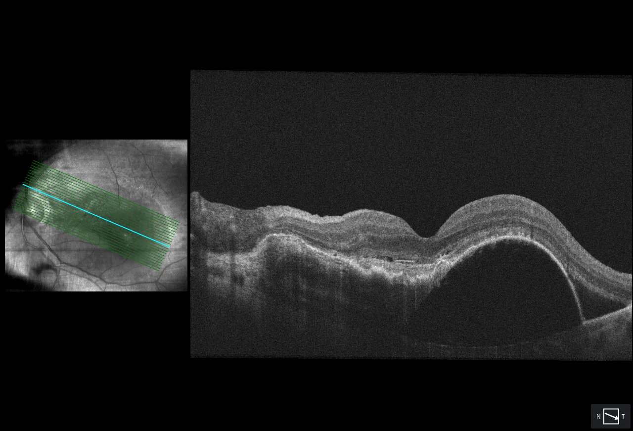
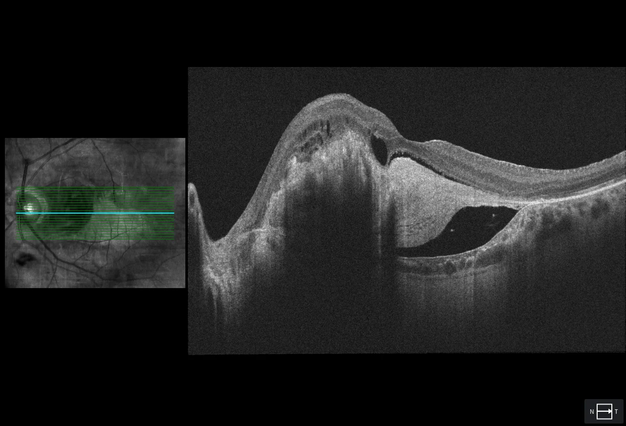
A: DEP with fibrovascular and serous component
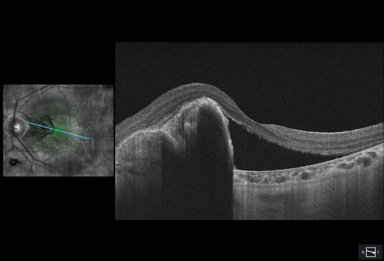
B3: RPE rupture with rolling towards the nasal
C3: onset of subretinal fibrosis after 6 months of follow-up
D3: severe subretinal fibrosis after 2 years of follow-up
Description
78-year-old woman with vision loss in left eye
MCVA OD 20/25 LE 20/100
Type 1 macular neovascularization. OCT shows a large PED with dual components, fibrovascular and serous, as well as perilesional subretinal fluid (A).
After 1 intravitreal injection of anti-VEGF, a tear of the RPE with rolling of the same towards the nasal is observed (B).
VA remains at 20/100 for a few months, but progression to subretinal fibrosis leaves poor hand movement VA after 6 months (C) and 2 years of follow-up (D).