Ampiginous Choroiditis
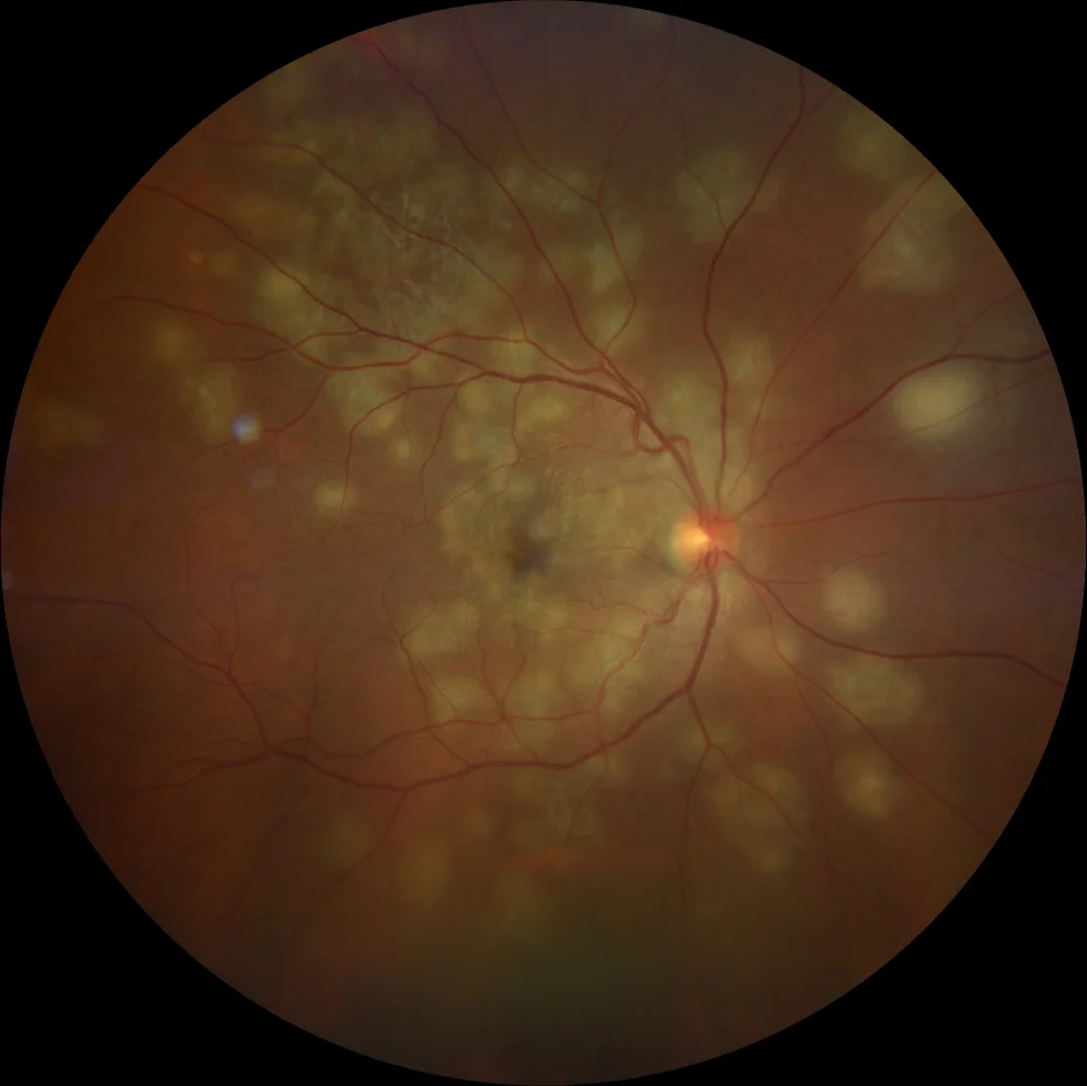
Color fundusography (Clarus 500, Carl Zeiss Meditec ASG, Jena, Germany) of the right eye in the acute phase and after treatment, respectively
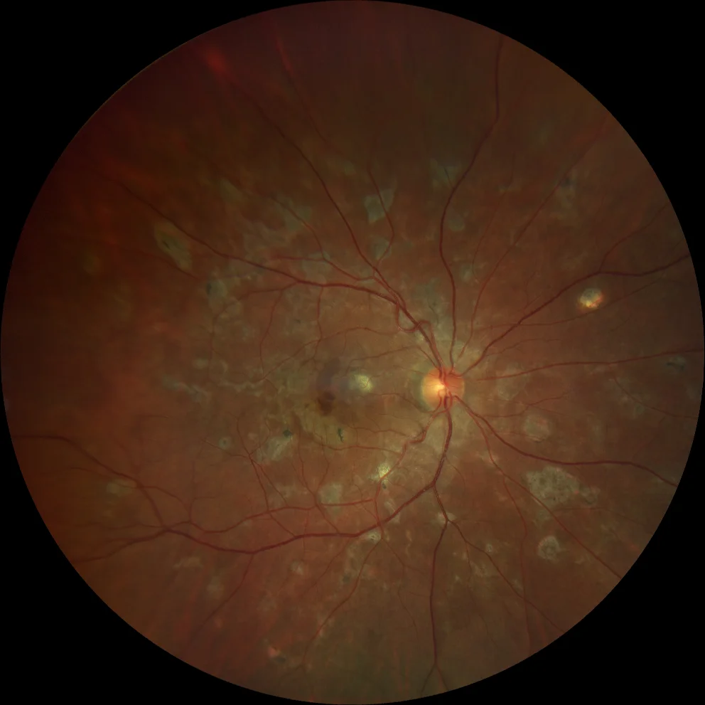
Color fundusography (Clarus 500, Carl Zeiss Meditec ASG, Jena, Germany) of the right eye in the acute phase and after treatment, respectively
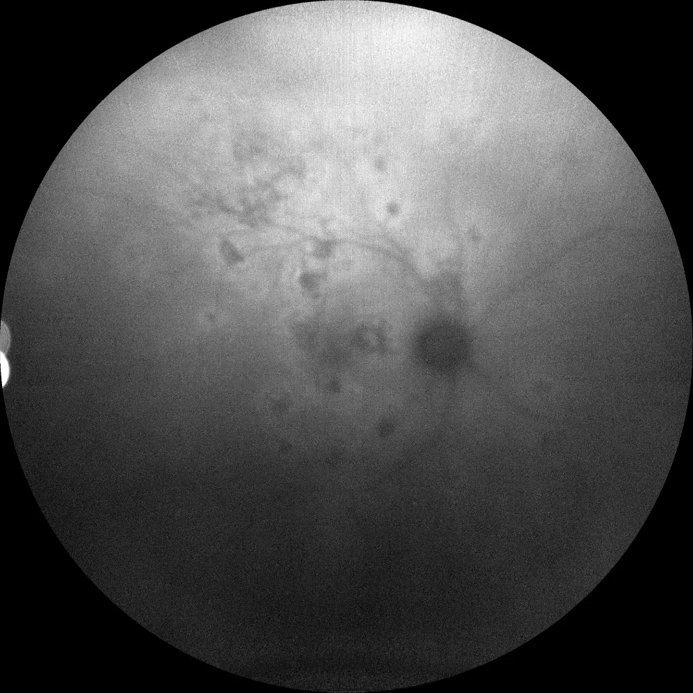
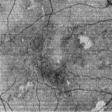
Autofluorescence (Clarus 500, Carl Zeiss Meditec ASG, Jena, Germany) of the right eye at diagnosis, at one month and after completion of treatment. It can be seen that the lesions are initially hyperautofluorescent with a hypoautofluorescent centre, and lose hyperautofluorescence as disease activity subsides. The presence of the hyperautofluorescent halo around the lesions indicates that the disease is still active.
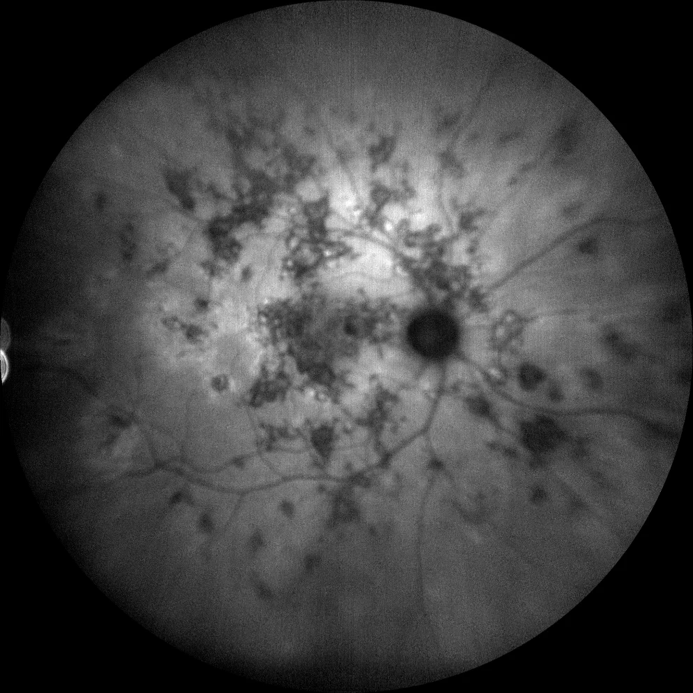
Autofluorescence (Clarus 500, Carl Zeiss Meditec ASG, Jena, Germany) of the right eye at diagnosis, at one month and after completion of treatment. It can be seen that the lesions are initially hyperautofluorescent with a hypoautofluorescent centre, and lose hyperautofluorescence as disease activity subsides. The presence of the hyperautofluorescent halo around the lesions indicates that the disease is still active.
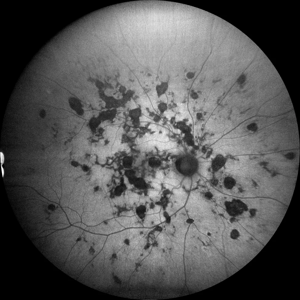
Autofluorescence (Clarus 500, Carl Zeiss Meditec ASG, Jena, Germany) of the right eye at diagnosis, at one month and after completion of treatment. It can be seen that the lesions are initially hyperautofluorescent with a hypoautofluorescent centre, and lose hyperautofluorescence as disease activity subsides. The presence of the hyperautofluorescent halo around the lesions indicates that the disease is still active.
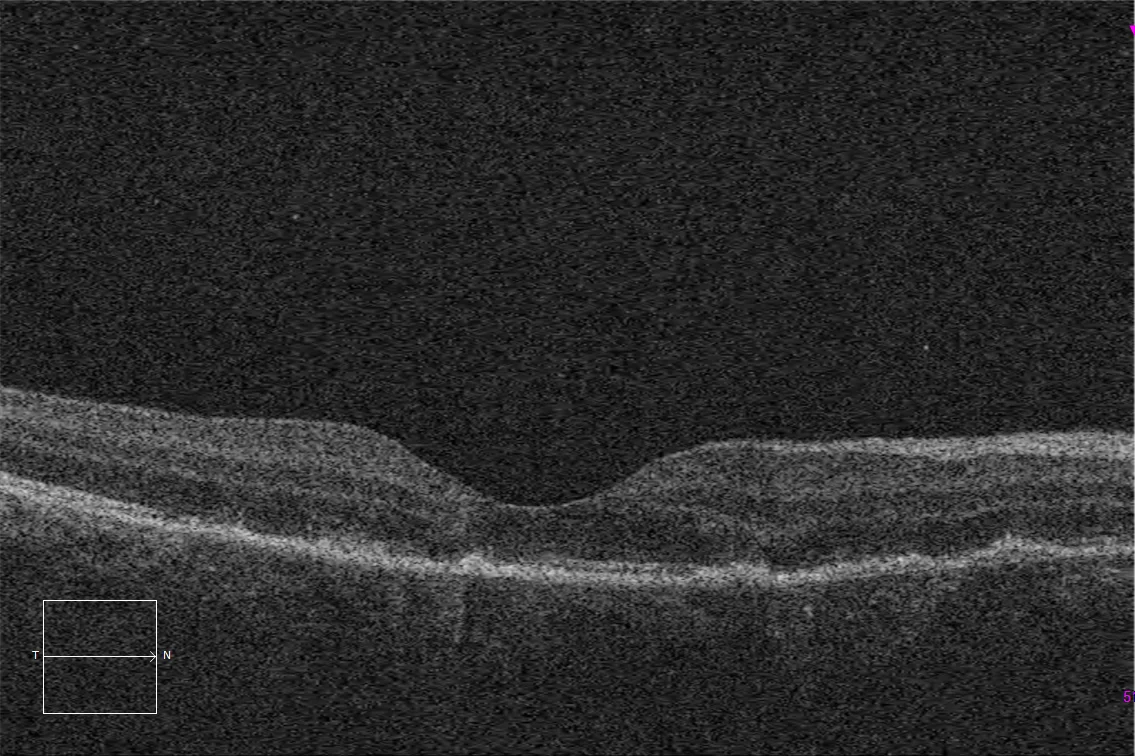
Optical coherence tomography (Cirrus 5000, Carl Zeiss Meditec ASG, Jena, Germany) on the fovea of the right eye at diagnosis, showing loss of the ellipsoid layer in the areas affected by the lesions, together with accumulations of hyperrefringent material.
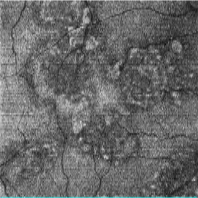
En face image of the macular optical coherence tomography (Cirrus 5000, Carl Zeiss Meditec ASG, Jena, Germany) of the right eye at the level of the ellipsoid, showing the loss of the external segments of the photoreceptors at the edges of the lesions, and the accumulations of hyperrefringent material in the central areas.
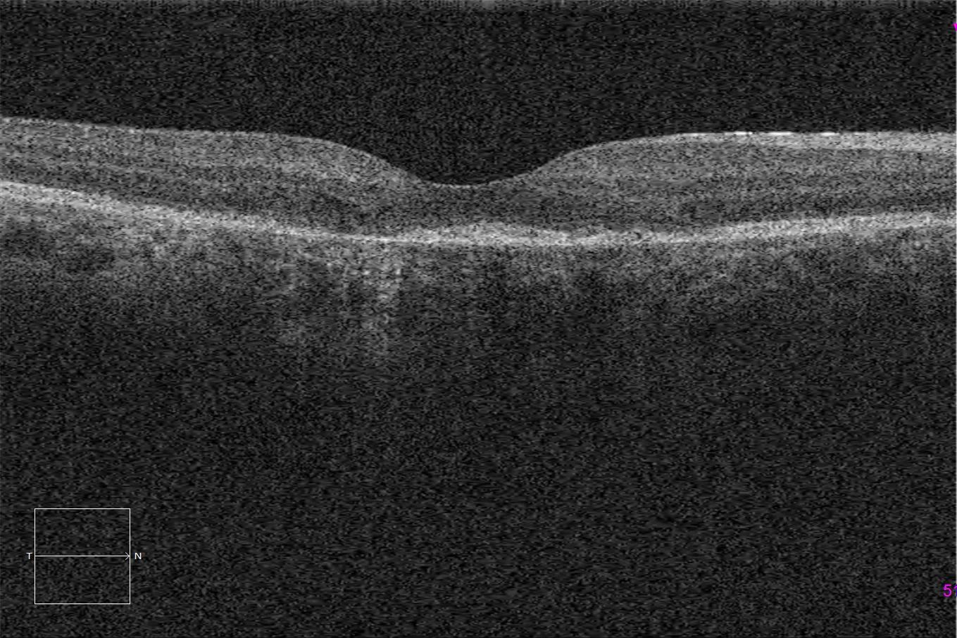
Optical coherence tomography (Cirrus 5000, Carl Zeiss Meditec ASG, Jena, Germany) over the fovea of the right eye after treatment, showing partial improvement of the ellipsoid layer.
En face image of the macular optical coherence tomography (Cirrus 5000, Carl Zeiss Meditec ASG, Jena, Germany) of the right eye at the level of the ellipsoid, showing the partial improvement of this layer after treatment.
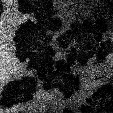
Optical coherence tomography - angiography (Cirrus 5000, Carl Zeiss Meditec ASG, Jena, Germany) at the level of the choriocapillaris, showing an absence of flow at the level of the lesions.
Description
Ampiginous choroiditis (or relentless placoid chorioretinitis) is a rare bilateral posterior uveitis affecting the retinal pigment epithelium (RPE) and the choroid. It presents with a clinical picture that is in the spectrum of acute posterior multifocal placoid pigment epitheliopathy (APMPEE), although in this case the involvement may progressively extend towards the periphery.
It affects men and women equally, between the second and sixth decades of life. It has a chronic, atypical and recurrent course. It is of unknown etiology, but it is postulated that there may be a possible viral, bacterial or autoimmune trigger. In the case of our patient, he was diagnosed with cutaneous tuberculosis concomitant with the ophthalmological picture. This form of presentation of ocular tuberculosis has been described between 9 and 12% of cases of ocular tuberculosis with choroidal involvement according to the COTS-1 study. The symptoms include rapidly appearing painless blurred vision, as well as paracentral scotomas, photopsia, metamorphopsia and myodesopsia. In the fundus we find multiple creamy and white lesions in the RPE, which end up progressing to chorioretinal scars. In autofluorescence, acute lesions are hyperfluorescent, while chronic ones are hypofluorescent. In optical coherence tomography we can find detachment of the RPE, as well as hyperreflectivity of the outer layers of the retina with loss of the ellipsoids in the acute phase of the disease, which can be reversed with appropriate treatment.
Comments
Despite the chronic course of the disease, the response to treatment is usually good if the condition is diagnosed and treated in time. Treatment may require corticosteroids, immunosuppressants and, in cases of infectious etiology such as this, specific antimicrobial treatment.Indication
A 40-year-old male presented with central blurred vision along with metamorphopsia in the right eye that had been developing for 4 days.