Birdshot chorioretinopathy
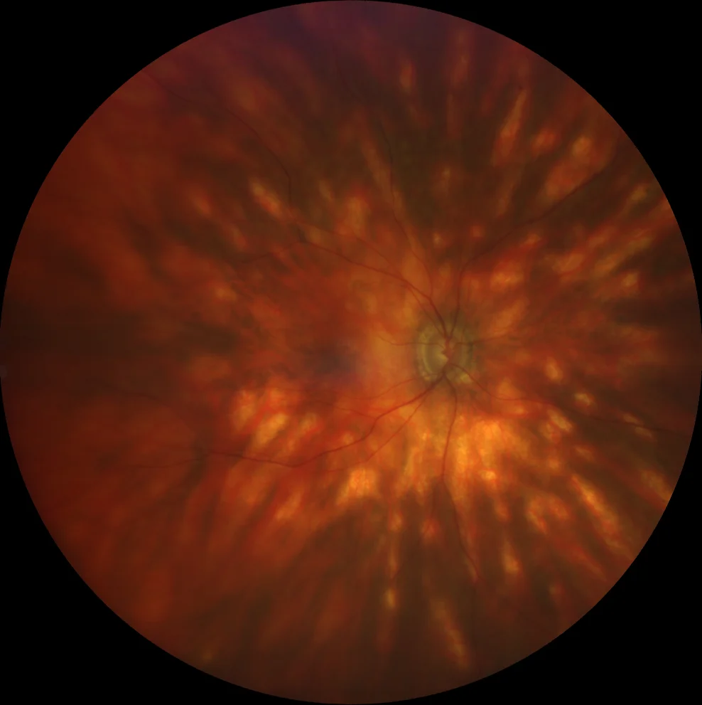
A and B. Color fundus photographs (Clarus 500, Carl Zeiss Meditec ASG, Jena, Germany) of the right and left eyes showing multiple hypopigmented choroidal lesions arranged radially around the optic disc.

A and B. Color fundus photographs (Clarus 500, Carl Zeiss Meditec ASG, Jena, Germany) of the right and left eyes showing multiple hypopigmented choroidal lesions arranged radially around the optic disc.
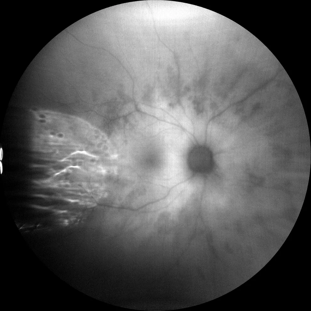
C and D. Autofluorescence (Clarus 500, Carl Zeiss Meditec ASG, Jena, Germany) of the right and left eyes showing hypoautofluorescence over the choroidal lesions.
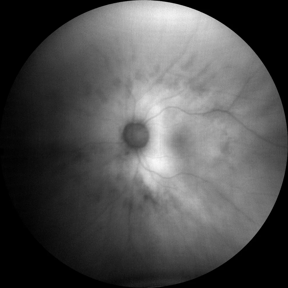
C and D. Autofluorescence (Clarus 500, Carl Zeiss Meditec ASG, Jena, Germany) of the right and left eyes showing hypoautofluorescence over the choroidal lesions.
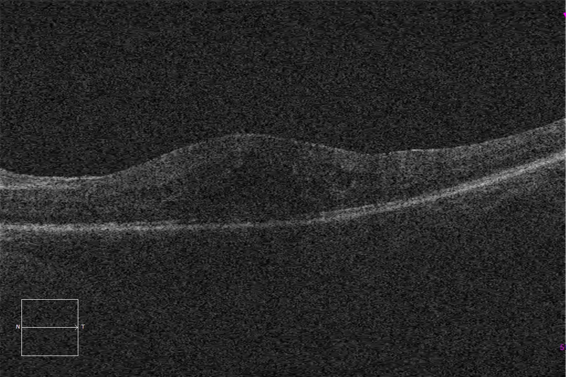
E and F. Macular optical coherence tomography (Cirrus 5000, Carl Zeiss Meditec ASG, Jena, Germany) of both eyes, showing cystoid macular edema, with inflammatory subfoveal neurosensory detachment in the right eye.
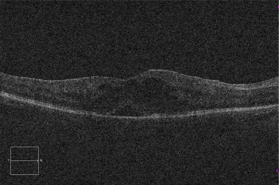
E and F. Macular optical coherence tomography (Cirrus 5000, Carl Zeiss Meditec ASG, Jena, Germany) of both eyes, showing cystoid macular edema, with inflammatory subfoveal neurosensory detachment in the right eye.
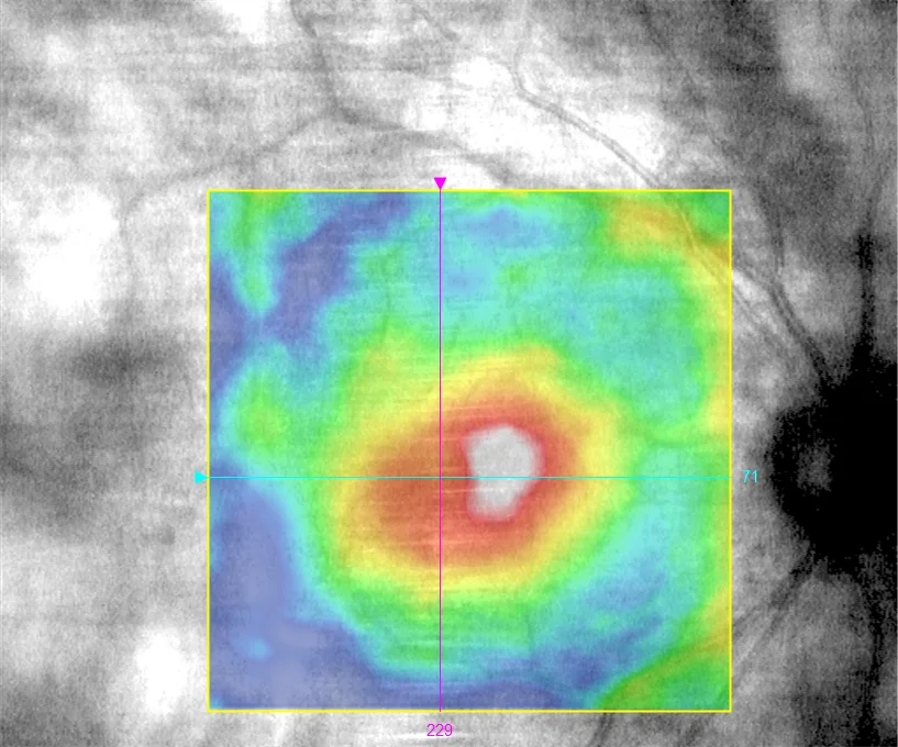
G and H. Color maps of macular retinal thickness measured by OCT (Cirrus 5000, Carl Zeiss Meditec ASG, Jena, Germany) of both eyes, showing bilateral macular edema.
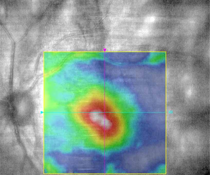
G and H. Color maps of macular retinal thickness measured by OCT (Cirrus 5000, Carl Zeiss Meditec ASG, Jena, Germany) of both eyes, showing bilateral macular edema.
Description
Birdshot chorioretinopathy is a rare disease, most common in women in northern Europe from the fifth decade of life onwards. There is no clear relationship with systemic diseases, but it is strongly associated with the HLA-A29 haplotype. The most characteristic finding in the fundus is the presence of hypopigmented lesions measuring 50 – 1500 μm, multifocal, ovoid, at the level of the choroid and retinal pigment epithelium that, over time, become depigmented. The radial distribution in the nasal sector is characteristic, starting at the optic nerve and following the choroidal vessels towards the periphery. Autofluorescence shows hypoautofluorescent lesions that may be more numerous than those observed in the fundus or color retinography. Optical coherence tomography (OCT) may reveal localized or diffuse loss of photoreceptors (internal-external segment line or ellipsoid zone) as well as macular edema. Choroidal thickness observed in OCT EDI may be increased in early stages and decreased in advanced disease.