Branch retinal arterial occlusion
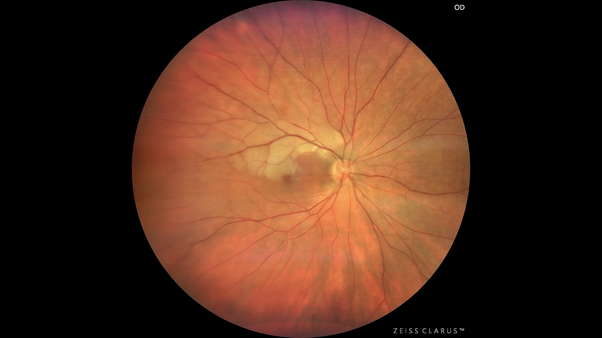
Color retinography showing a yellowish-white pallor over the upper temporal arch.
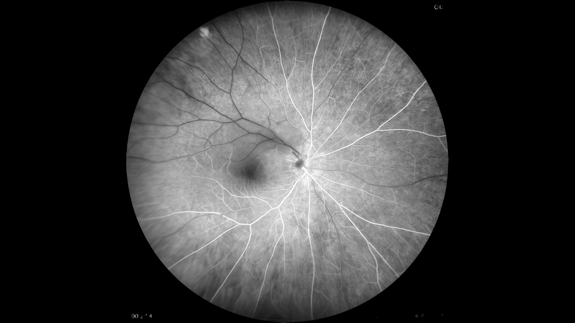
Fluorescein angiography: showing arterial occlusion in the territory of the superior temporal branch, together with the corresponding delay in venous filling of its homologous vein.
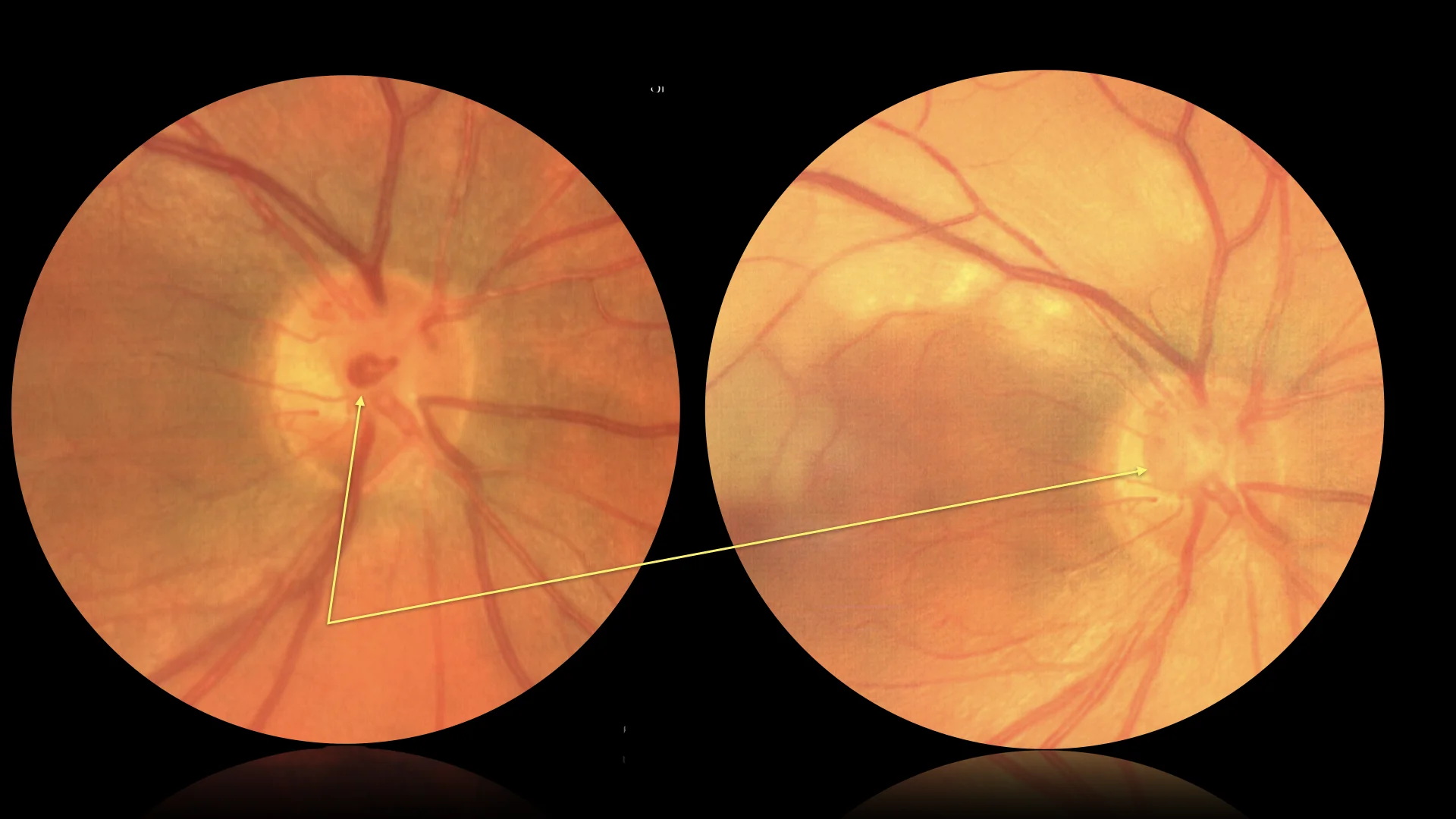
Montage of 4 pre and post occlusion images, in which the closure of the loop and the arterial blood supply of the upper temporal arch can be observed in detail.
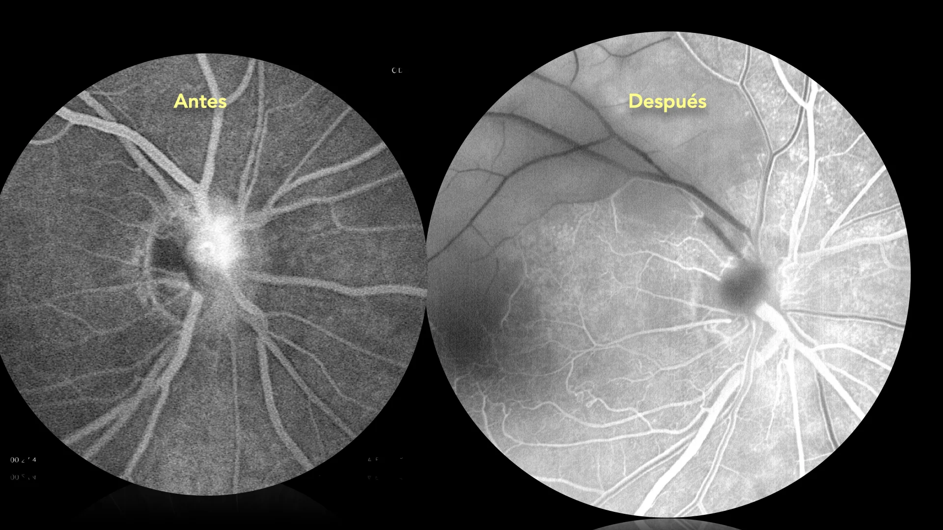
Montage of 4 pre and post occlusion images, in which the closure of the loop and the arterial blood supply of the upper temporal arch can be observed in detail.
Description
Branch retinal artery occlusion (BRAO) is an ophthalmologic emergency characterized by blockage of a minor retinal artery, resulting in sudden, focal vision loss. This event is usually caused by an embolus obstructing blood flow to a portion of the retina. On retinography, RRAO is visualized as areas of retinal whitening that correspond to the ischemic zone affected by the lack of blood flow. In addition, narrowed arterioles are seen and, depending on the location of the occlusion, a cherry red spot reflex may appear in the fovea if the macular area is involved. These signs are indicative of the severity and extent of the arterial blockage.